
We present recent clinical and diagnostic advances in spinal cord imaging. Because of the overlap of different pathologic entities, good knowledge of clinical information is necessary. Degenerative diseases of the spine can sometimes be misleading when the question of a possible tumor rises. The essentials of spinal cord tumors are discussed.
In patients with suspicion of demyelinating disease, the following considerations are of importance: multiple sclerosis (MS) should be differentiated from neuromyelitis optica spectrum disorder (NMOSD), and AQP4 and MOG-antibodies should be searched for. There is a growing body of evidence that there is an overlap between NMOSD and acute demyelinating encephalomyelitis (ADEM).
Acute transverse myelitis is often a diagnosis of exclusion after viral myelitis or autoimmune causes have been excluded. The involvement of specific areas can give an indication for the diagnosis, e.g., area postrema or conus medullaris. New MRI equipment enables more robust diffusion-weighted imaging (DWI) of the spine, and DWI is becoming a valuable tool for radiologists (Figure 1a–c). Finally, some examples of pathology were seen on CT that illustrate that MRI is not the unique imaging examination of choice in spinal cord diseases.
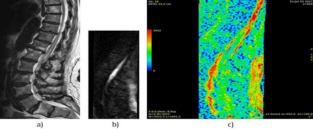
Figure 1
81y old woman who woke up in the morning with paraparesis of the legs, Hyperintense lesion of the conus medullaris on Sagittal FSE T2 image (a) with acute oedema of the conus of Focus DWI-B600 (b and c).
Competing Interests
The author has no competing interests to declare.