
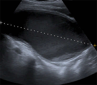
Figure 1
Sagittal US-image showing a large hypogastric mass with hyporeflective content and a small amount of hyperreflective material posteriorly.
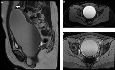
Figure 2
MRI: A. Sagittal T2-weighted image showing a large cystic mass with T2 hyper- to iso-intense content. Arrow = uterine cervix. B. Axial T1-weighted image showing a large cystic mass with T1 hyperintense content. C. Axial T2*-weighted image showing susceptibility artifacts posteriorly in the lesion.