
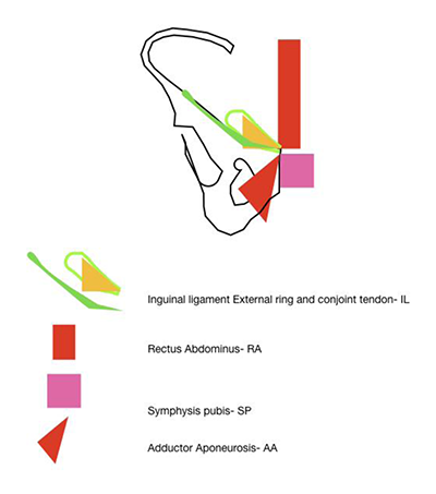
Figure 1
Structures attached to the pubic tubercle.
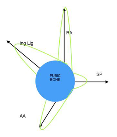
Figure 2
Vectors on the pubis as a clock-face.
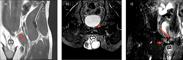
Figure 3
a) 30 year old footballer with left groin pain. Coronal oblique high resolution image through the medial aspect of the inguinal ligament adjacent to the pubic enthesis demonstrates a cord-like medial aspect of the inguinal ligament (red arrow). b) 30 year old footballer with left groin pain. Angled oblique axial inlet High resolution T2 fat suppressed images through the superior aspect of the pubic bone at the site of the inguinal ligament insertion demonstrates BMO of the left pubic bone (red arrowhead) c) 30 year old footballer with left groin pain. Coronal oblique High resolution T2 fat suppressed images through the adductor insertion on the right and pubic tubercle on the left, demonstrates site specific BMO of the left pubic bone at the inguinal entheses (long red arrow) and a right adductor cleft with associated site specific BMO (short red arrow).