
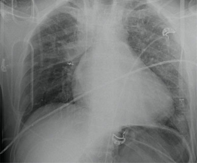
Figure 1
Multiple left sided rib fractures without pneumothorax and blurry consolidations in both lungs, probably lung contusions.
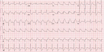
Figure 2
ECG on admission shows sinus tachycardia (109 bpm) and the presence of an ST segment elevation inferior. The ST elevation in lead III is more pronounced than in lead II, which suggests an occlusion of the RCA. There is also an ST elevation in leads V1–V3, with more important ST elevation in lead V1 than V2, suggestive of concomitant right ventricular infarction.
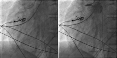
Figure 3
Coronary angiography. Left side image showing guide in the RCA. On the right-side image during injection of iodized contrast showing backflow in the aorta and no enhancement of the RCA compatible with complete occlusion.
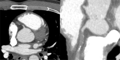
Figure 4
Spiral acquisition trauma CT of the thorax and abdomen after intravenous injection of 100 ml iodized contrast. Focus on the heart showing dissection and complete occlusion of the RCA. Left image: axial view, right image: curved view.