
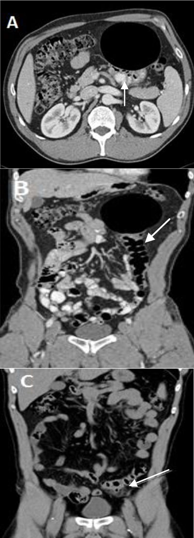
Figure 1
(A) and (B) Contrast-enhanced CT scan showing a 10.4 × 8.1 cm smooth-walled well-defined cystic air-filled lesion adjacent to the sigmoid (arrow) lifted in the left hypochondrium. (C) Unenhanced CT scan of the abdomen from one year earlier showed sigmoid diverticulitis (arrow) located in the left iliac fossa and without the cystic mass.
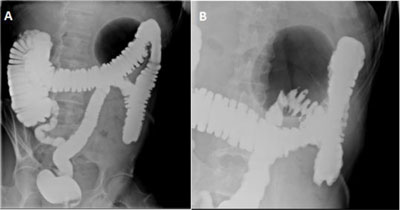
Figure 2
Gastrografin enema in standing position shows (A) a well-defined large gas-filled cavity of 9.7 cm in the left hypochondrium with neither air-fluid level nor extravasation of the contrast agent (balloon sign) and (B) focalized diverticular disease.

Figure 3
Photomicrograph (hematoxylin-eosin stain) shows the diverticulum consisting of fibrosclerotic wall colonized by mixed inflammatory elements with a complete destruction of the endodiverticular mucosal layer (McNutt type 2).