
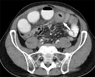
Figure 1
Axial view of contrast-enhanced CT reveals circumferential small bowel wall distention (arrow) and engorged mesenteric vessels (*).
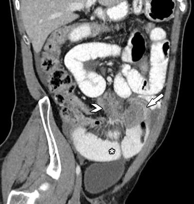
Figure 2
Oblique coronal view of contrast-enhanced CT shows the close relation between the mesenteric mass (arrowhead) and its extension towards the small bowel (arrow). Note the distended small bowel loops (*).
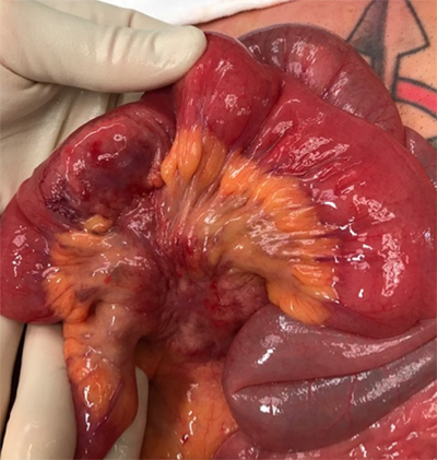
Figure 3
Peroperative image reveals the small bowel wall invasion and mesenteric mass.
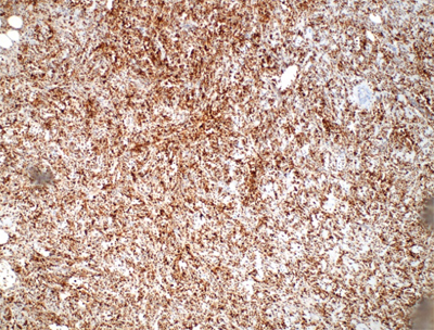
Figure 4
Immunohistological staining reveals highly positive lysozyme markers.