

Figure 1
Contrast-enhanced axial CT of the brain shows fusiform and contrast-enhancing thickening of the left lateral and medial rectus muscles.
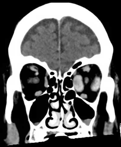
Figure 2
Coronal reconstructions of contrast-enhanced brain CT show fusiform thickening of the left medial and lateral rectus muscles as well as slight thickening of the left superior rectus muscle.
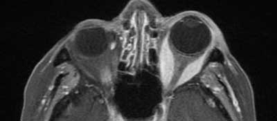
Figure 3
Contrast-enhanced fat suppressed axial T1-weighted MR-images of the orbit confirm fusiform thickening of the left medial and lateral muscles. No extension of intra-orbital changes into the cavernous sinus is seen.