
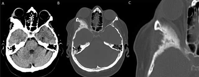
Figure 1
Non-contrast enhanced axial CT of the brain with soft tissue (A) and bony window reconstructions (B) shows an expansile sclerotic lesion in the right sphenoid wing. Involvement of the right lateral orbital wall leads to narrowing of the orbital space and secondary proptosis. A zoomed-in view in bony window (C) shows the irregularly marginated sclerotic expansion of the right lateral orbital wall in more detail.
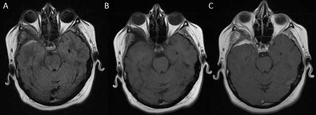
Figure 2
MRI of the brain with axial FLAIR (A) and unenhanced and contrast-enhanced axial T1-weighted images (B, C) shows right-sided anterotemporal FLAIR-hyperintense contrast-enhancing dural thickening abutting the bony sphenoid changes.