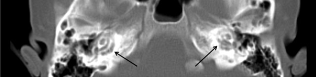
Have a personal or library account? Click to login
Temporal Bone Imaging Features in Osteogenesis Imperfecta Cover

Temporal Bone Imaging Features in Osteogenesis Imperfecta

Open Access
|Aug 2017

Figures & Tables

jbsr-101-1-1321-g1.jpg
Figure 1

Axial CT-scan shows bilateral lucencies in the pericochlear bony otic capsule.
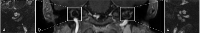
jbsr-101-1-1321-g2.jpg
Figure 2

Coronal reconstructions of contrast-enhanced 3D FFE-imaging demonstrates symmetric areas of contrast enhancement in the pericochlear regions (b) Hyperintense signal anomalies are seen around the basal turn of the cochlea in the right (a) and left temporal bones (c) on 3D balanced steady-state gradient echo-imaging.

Language: English
Published on: Aug 11, 2017
Published by: Ubiquity Press
In partnership with: Paradigm Publishing Services
Publication frequency: 1 issue per year

© 2017 Isabeau Hermie, Monique Horvath, Sofie Van Cauter, published by Ubiquity Press
This work is licensed under the Creative Commons Attribution 4.0 License.