
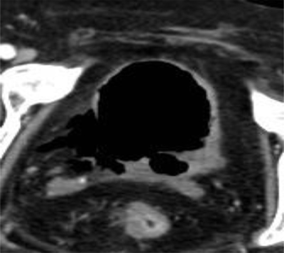
Figure 1
Axial unenhanced CT scan of the abdomen shows a disruption of the bladder wall at the right posterolateral side.
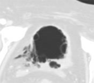
Figure 2
Same Axial unenhanced CT-scan image as above in Lung Window shows fibrotic tissue at the disrupton site. There are multiple air-filled submucosal blebs of the bladderwall suggesting an emphysematous cystitis.
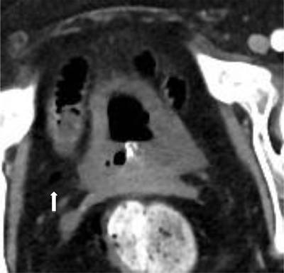
Figure 3
One-week later follow-up CT was performed, this showed a spontaneous healing of the bladder wall with only a small residual focus of free air in the Retzius space on the right (white arrow).