
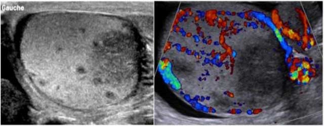
Figure 1
Grayscale and color Doppler of the left testis demonstrate hypoechoic avascular area of the hilum; the remaining testicular and epididymal parenchyma are more hyperechoic with flow detection.
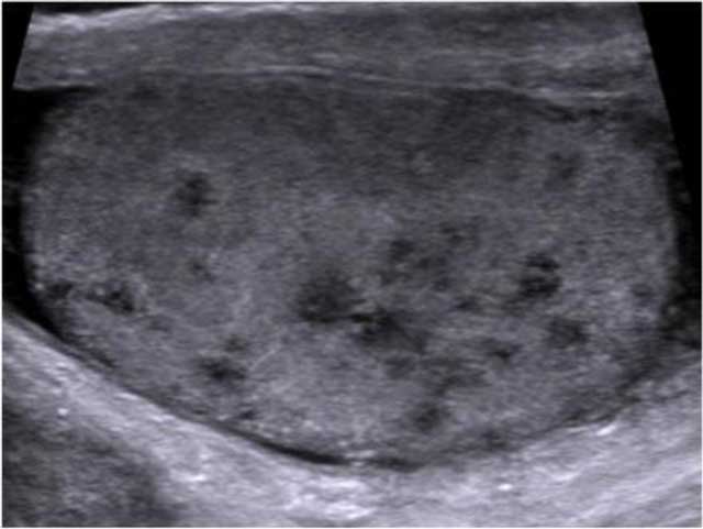
Figure 2
Longitudinal US image of the left testis shows multiple small hypoechoic nodules (< 2–3 mm) corresponding to TBC granulomas in a miliary pattern.
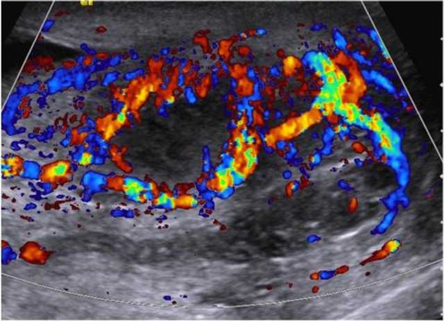
Figure 3
Longitudinal color Doppler image shows hypoechoic abscess of the hilum extending to the epididymis with increased flow signals in peripheral portion.
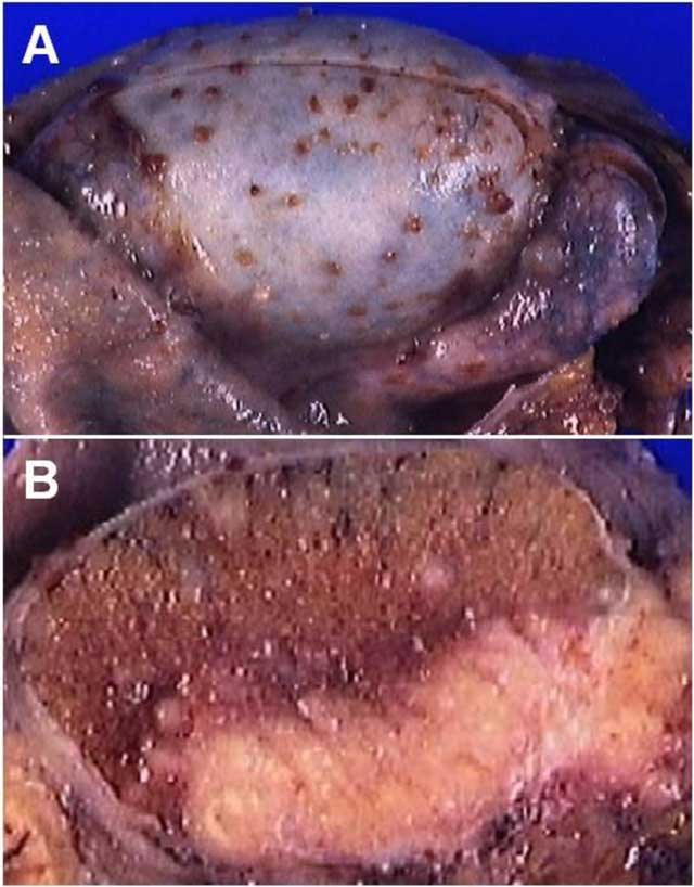
Figure 4
Histopathology specimens of the left testis. (A) Chirurgical specimen of left orchidectomy with many brownish nodules at the tunica albuginea and epididymis. (B) Cross section of left testicle showing caseating necrosis of the hilum and the epididymal head with muliple white millimetric nodules.