
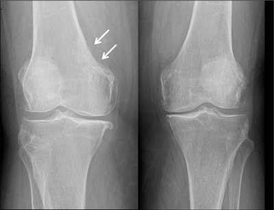
Figure 1
AP views of both knee joints: Obvious bilateral gonarthrosis. The erosive lesion in the medial cortex of distal metaphysis of right femur and associated discrete subcortical sclerosis (arrows) can be easily missed. Compare with the smoothly outlined medial cortex of left femur.

Figure 2
Skeletal scintigraphy – corresponding metabolically active lesion in the right femur (arrow) and multiple other widespread skeletal metastases.
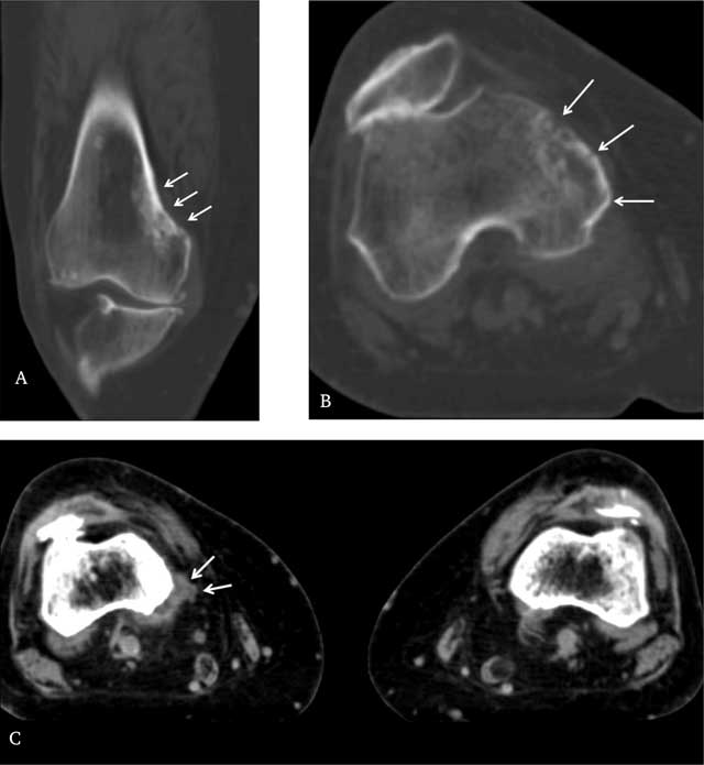
Figure 3
A. Coronal reconstructed CT image and B. axial CT image through distal right femur in bone window show the extent of the cortical metastases and underlying sclerosis more clearly (arrows). C. Axial CT scan through distal femur in soft tissue window shows a discrete associated soft tissue mass posteromedially on the right side, compared with the normal left side (arrows).