
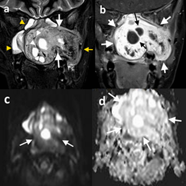
Figure 1
13-year-old male patient (patient 5), (a) coronal T2-weighted precontrast, (b) coronal T1-weighted postcontrast, (c) DWI, and (d) ADC map demonstrate a left-sided nasopharyngeal mass which enlarges the ipsilateral pterygopalatine fossa (a, white arrows) and extends into the temporal fossa (a, yellow arrow). The mass is hyperintense on T2-weighted image (a) and exhibits significant contrast enhancement (b). Diffusion-weighted images (c) showed no diffusion restriction, and the lesion has high signal intensity on the ADC map (d). The tumor demonstrates internal cystic components (b, black arrows) and signal-void regions (a, white arrowheads). Image a demonstrates inflammatory signal changes in maxillary and sphenoid sinuses (yellow arrowheads).
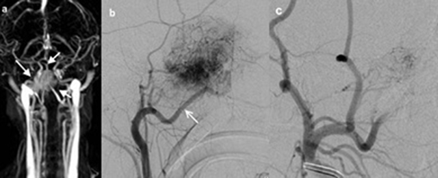
Figure 2
10-year-old male patient (patient 3), (a) TWIST-MR angiography of the patient showed bilobule hypervasculer mass on the right side (white arrows). (b) Selective right carotid artery angiography shows that JNA is supplied with the right internal maxillary artery (white arrow). (c) It is observed that the opacification of JNA mainly dissappeared in the angiography display obtained after the internal maxillary artery was embolized with microcoil.
Table 1
Magnetic Resonance İmaging Findings of the Patients.
| Patient No | 1 | 2 | 3 | 4 | 5 | 6 |
|---|---|---|---|---|---|---|
| Tumor size | 43 × 40 × 47 | 72 × 70 × 64 | 52 × 62 × 45 | 38 × 43 × 33 | 101 × 72 × 81 | 54 × 58 × 48 |
| Contrast enhancement | Intense | Intense | Intense | Intense | Intense | Intense |
| Flow-voids | + | + | + | + | + | + |
| T1WI | Hyperintense | Isointense | Iso-/Hyperintense | Isointense | Isointense | Iso-/Hypointense |
| T2WI | Hyperintense | Iso-/Hyperintense | Hyperintense | Iso-/Hyperintense | Hyperintense | Iso-/Hyperintense |
| DWI | – | – | – | NA | – | NA |
| ADC value (mm2/s) | 1,5 × 10⁻³ | 1,5 × 10⁻³ | 1,6 10–3 | NA | 1,7 × 10⁻³ | NA |
| Antral sign | + | – | – | + | – | + |
| Cystic component | – | + | + | – | + | + |
| Vascular compression | – | –* | – | – | – | – |
[i] NA Not available.
*Surrounds the internal carotid artery without compression.
Table 2
Extension Areas of the Tumors.
| Patient no. | 1 | 2 | 3 | 4 | 5 | 6 |
|---|---|---|---|---|---|---|
| Localization | Right | Right | Right | Right | Left | Right |
| Nasopharynx | + | + | + | + | + | + |
| Nasal cavity | + | + | + | + | + | + |
| Pterygopalatine fossa | + | + | + | + | + | + |
| Infratemporal fossa | – | + | + | + | + | + |
| Maxillary sinus | – | + | – | – | + | – |
| Ethmoid sinus | – | + | + | – | + | + |
| Sphenoid sinus | – | + | + | – | + | + |
| Cheek | – | + | – | – | + | + |
| Orbit | – | + | – | – | + | – |
| Cavernous sinus | – | + | – | – | + | + |
| Optic chiasm | – | + | – | – | – | – |
| Skull base erosion | – | + | + | – | + | + |
| Middle cranial fossa | – | + | + dural | – | + dural | – |
| Bone marrow edema | + | + | + | + | + | + |
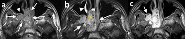
Figure 3
15-year-old male patient (patient 1), (a) axial T1-weighted precontrast, (b) axial T2-weighted precontrast, and (c) axial T1-weighted postcontrast MR images demonstrate a right-sided nasopharyngeal mass which enlarges the ipsilateral pterygopalatine fossa (a and b white arrows). The mass is isointense with the muscle on T1-weighted image (a) and hyperintense on T2-weighted image (b) and exhibits significant contrast enhancement (c). The posterior wall of the maxillary sinus demonstrates anterior bowing with resultant Holmann Miller sign (a, white arrowhead; c, arrow). The tumor demonstrates signal-void regions (b, yellow arrow) and inflammatory signal changes in maxillary sinuses (b, white arrowhead).
Table 3
Preoperative Stages of the Patients, According to Radkowski and Onerci Staging Systems.
| Patient no. | Radkowski | Onerci |
|---|---|---|
| 1 | IIB | II |
| 2 | IIIB | IV |
| 3 | IIIA | III |
| 4 | IIC | II |
| 5 | IIIA | IV |
| 6 | IIIA | IV |
Table 4
Summary of the Staging Systems.
| Radkowski et al. 1996 |
|---|
| IA Tumor limited to nasal cavity/nasopharynx IB Extension into one or more paranasal sinuses IIA Minimal extension into pterygopalatine fossa IIB Invasion of the pterygomaxillary fossa (with or without erosion of the orbital bones IIC Tumor extension into infratemporal fossa, with or without the involvement of the cheek or the pterygoid plates IIIA Erosion of the skull base, with minimal extension into cranial fossa IIIB Prominent intracranial extension, with or without invasion of the cavernous sinus |
| Onerci et al. 2006 |
| I Minimal extension into nasal cavity, nasopharynx, ethmoid-sphenoid sinuses, or pterygomaxillary fossa II Extensive invasion of maxillary sinuses and the pterygomaxillary fossa, extension into anterior cranial fossa and limited extension into infratemporal fossa III Marrow involvement of the body and the greater wing of sphenoid bone and base of the pterygoid; extensive involvement of the infratemporal fossa, pterygoid plate, or orbital region; obliteration of cavernous sinus IV Intracranial extension between the pituitary and ICA, tumor located lateral to ICA, extension into middle cranial fossa |