
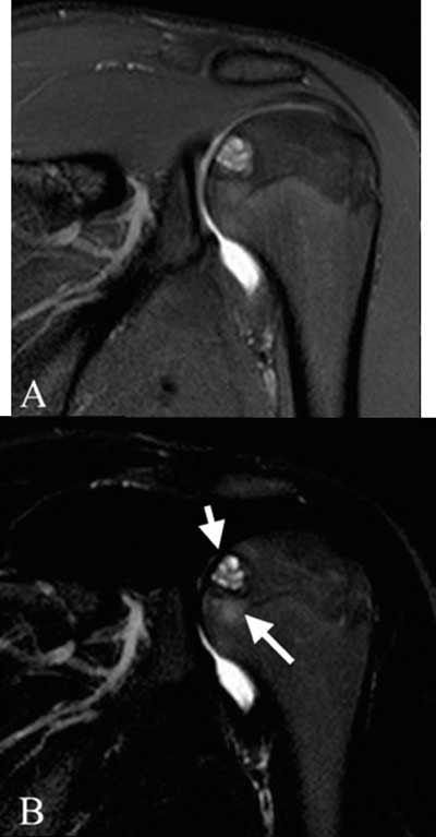
Figure 1
Coronal dual TSE FS images of the left shoulder. (A): proton density weighted image: well described lesion in the proximal humeral epiphysis without disruption of the subchondral bone plate, (B): T2-weighted: central high signal intensity (short arrow), limited bone marrow edema (long arrow).
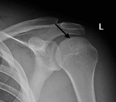
Figure 2
Radiograph of the left shoulder, AP view: a rounded lytic lesion is seen in the epiphysis of the humeral head, with sharp borders and a thin sclerotic rim (arrow).
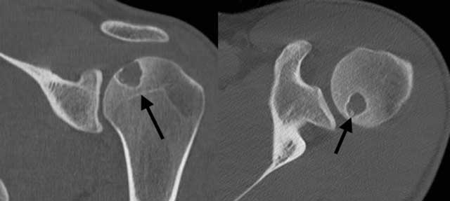
Figure 3
CT of the left shoulder. (A) Coronal reformatted image: the lesion does not reach the growth plate, which is already closed (arrow); (B) Axial plane image: a few small dense fragments can be seen in the lytic lesion (arrow): calcifications, confirming the presence of a chondroid matrix.
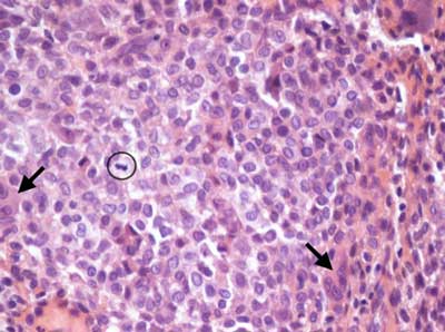
Figure 4
Histological image (HE staining, enlargement 40X) of the resected chondroblastoma: compact rounded tumor cells with bean-shaped nuclei, giant cells (arrows), a mitotic figure (circle) and chondroid matrix (red).