
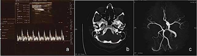
Figure 1
Ultrasound and Color Doppler images reveal right ICA agenesis and externalization flow pattern of right CCA (a); an axial CT image at bone window reveals absent right bony carotid canal (b); MR angiography image shows right ICA agenesis and anomalous origin of ophthalmic artery (c) in Case 1.

Figure 2
An axial CT image at bone window reveals absent right bony carotid canal (a); cranial CT image reveals hypodense lesion at right subthalamic region about 1 cm in diameter (b) in Case 2.
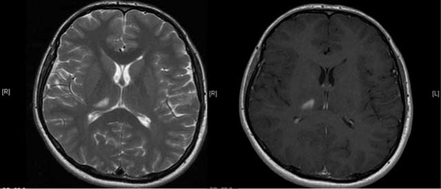
Figure 3
An axial T2W and enhanced T1W images show lesion consistent with subacute infarction (Case 2).