
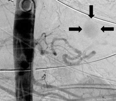
Figure 1
Abdominal aortic angiogram: the 2.5 cm large aneurysm of the distal splenic artery (arrows).
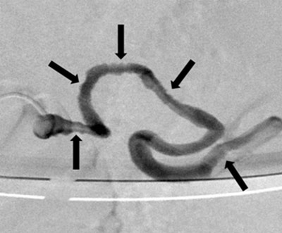
Figure 2a
Selective splenic artery opacification: the multiple parietal irregularities (arrows).
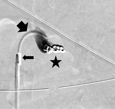
Figure 2b
Postostial occlusion of the splenic artery: the 5F support catheter (thin arrow), the 3F microcatheter (thick arrow), and the microcoils (star).
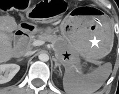
Figure 3
Axial contrast-enhanced CT image: the pancreatic neoplasm (black star) and the almost entirely clotted gastric lumen (white star).
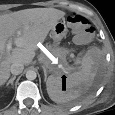
Figure 4a
Enhanced CT, a few days later: recurrent hemorrhage (white arrow) arising retrogradely from the distal splenic artery (black arrow).
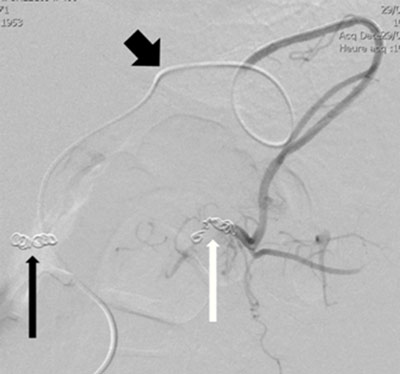
Figure 4b
Microcoils embolization of the distal splenic artery (white arrow) through superselective catheterism of the gastroepiploic arteries (thick black arrow); the thin black arrow indicates the previous ostial coiling.