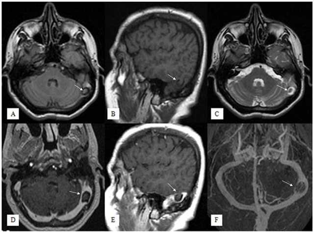
Have a personal or library account? Click to login
Temporal Lobe Parenchyma Herniation into the Transverse Sinus: MRI Findings in a Case Cover

Temporal Lobe Parenchyma Herniation into the Transverse Sinus: MRI Findings in a Case

Open Access
|Jan 2016

Figures & Tables

figures/Fig01_web.jpg
Figure 1

Fluid Attenuated Inversion Recovery axial (A), T1-weighted sagittal (B), T2-weighted axial (C), contrast-enhanced T1-weighted axial (D) and sagittal (E) images show a small herniation of temporal lobe parenchyma with surrounding CSF into left transverse sinus, that was isointense to brain parenchyma on all sequences (arrows). No pathological enhancement is seen but the brain herniation sac is causing moderate stenosis in the left transverse sinus. On venography imaging (F), there was left transverse sinus stenosis but no venous thrombosis.

Language: English
Published on: Jan 29, 2016
Published by: Ubiquity Press
In partnership with: Paradigm Publishing Services
Publication frequency: 1 issue per year

© 2016 Elçin Aydın, Hasan Yerli, Esin Gezmiş, published by Ubiquity Press
This work is licensed under the Creative Commons Attribution 4.0 License.