
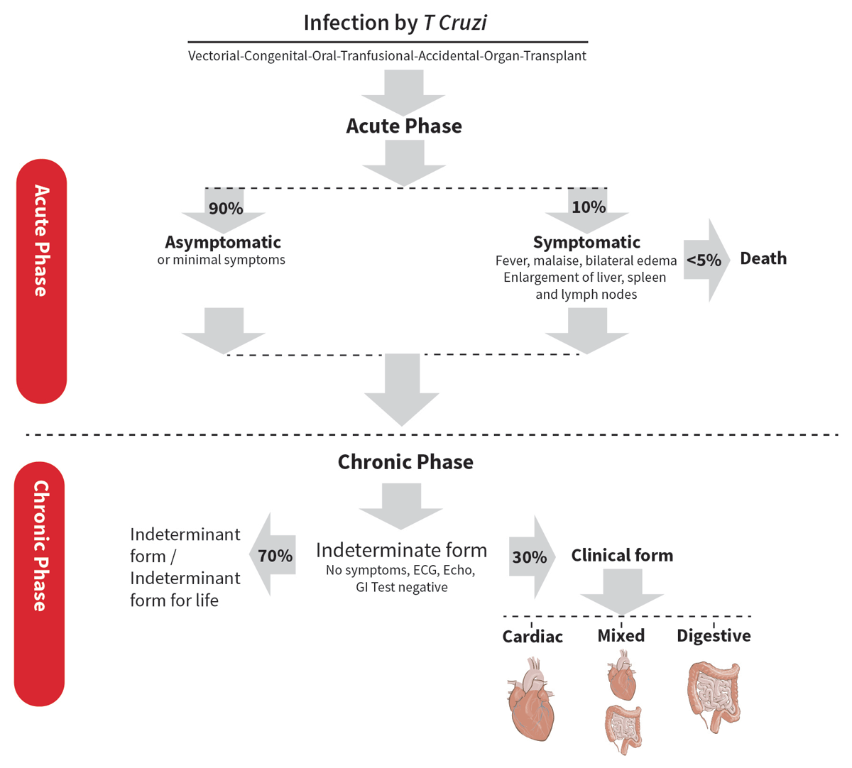
Figure 1
Phases of Chagas disease [1].
Table 1
Recommendations on indications for performing echocardiography.
| INDICATION | APPROPRIATE (A), INAPPROPRIATE (I) OR UNDETERMINED (U) |
|---|---|
| Perform an echocardiogram for the diagnosis of Chagas disease | I |
| Perform a transthoracic echocardiogram to assess cardiac involvement in a patient diagnosed with Chagas disease | A |
| In areas with limited accessibility to health centers, it would be useful to perform a handheld echocardiogram to initially assess cardiac involvement | U |
| In rural areas with limited access to health centers, a POCUS protocol could be proposed to assess wall motion disorders, the presence of ventricular aneurysms, and left ventricular ejection fraction in patients with Chagas disease to determine cardiac involvement | U |
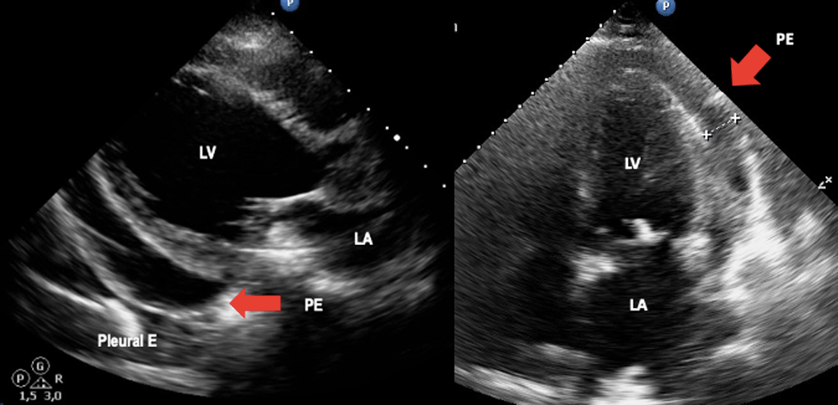
Figure 2
Acute Chagas disease. Arrows indicate pericardial effusion in heart failure due to Chagas disease. Image: Mariana Corneli. Reproduced with permission of the photographer.
Table 2
Chagas disease staging.
| ACUTE PHASE | CHRONIC PHASE | ||||
|---|---|---|---|---|---|
| INDETERMINATE FORM | CHAGAS CARDIOMYOPATHY | ||||
| A | B1 | CHAGAS DILATED CARDIOMYOPATHY/HEART FAILURE | |||
| B2 | C | D | |||
| Myocardial wall motion and/or pericardial effusion | Patients at risk for developing HF. They have positive serology, neither structural cardiomyopathy on ECG/echo nor HF symptoms. | Patients with structural cardiomyopathy, evident on ECG or echocardiographic abnormalities, but with normal or mildly depressed global LV function and neither current nor previous HF symptoms. | Patients with structural cardiomyopathy characterized by global LV dysfunction (LVEF > 40% < 50%) and neither current nor previous signs and symptoms of HF | Patients with ventricular dysfunction (LVEF < 40%) or V aneurism and current or previous symptoms of HF (NYHA FC I, II, III, or IV) | Patients with refractory symptoms of HF at rest despite optimized clinical treatment requiring specialized interventions |
Table 3
Recommendations for use of echocardiography in evaluation of the left ventricle.
| PARAMETER OR INDICATION | APPROPRIATE (A), INAPPROPRIATE (I) OR UNDETERMINED (U) |
|---|---|
| 2D Echo Assessment of LV Dimensions | A |
| Assessment by calculation of LVEF through the biplane method of disks (the Simpson rule) | A |
| LV evaluation in the presence of ventricular aneurysm by 3D echo if this methodology is available | A |
| Identify segmental wall motion disorders through 2D echo | A |
| Evaluation of LV systolic function by 2D echo in the presence of apical aneurysm and 3D echo available | I |
| Evaluation of global longitudinal deformity of the LV in the absence of motility disorders and preserved FEY in patients with suspected indeterminate form | A |
| It is recommended to evaluate the presence of ventricular dyssynchrony in patients with Chagas disease and impaired LVEF | A |
| Use of contrast echo in case of poor ultrasound window or in case of suspected left ventricular aneurysm if it is available | A |
| In a patient with Chagas disease and an echocardiogram without abnormalities, perform a stress echo with dobutamine to unmask ventricular dysfunction. | U |
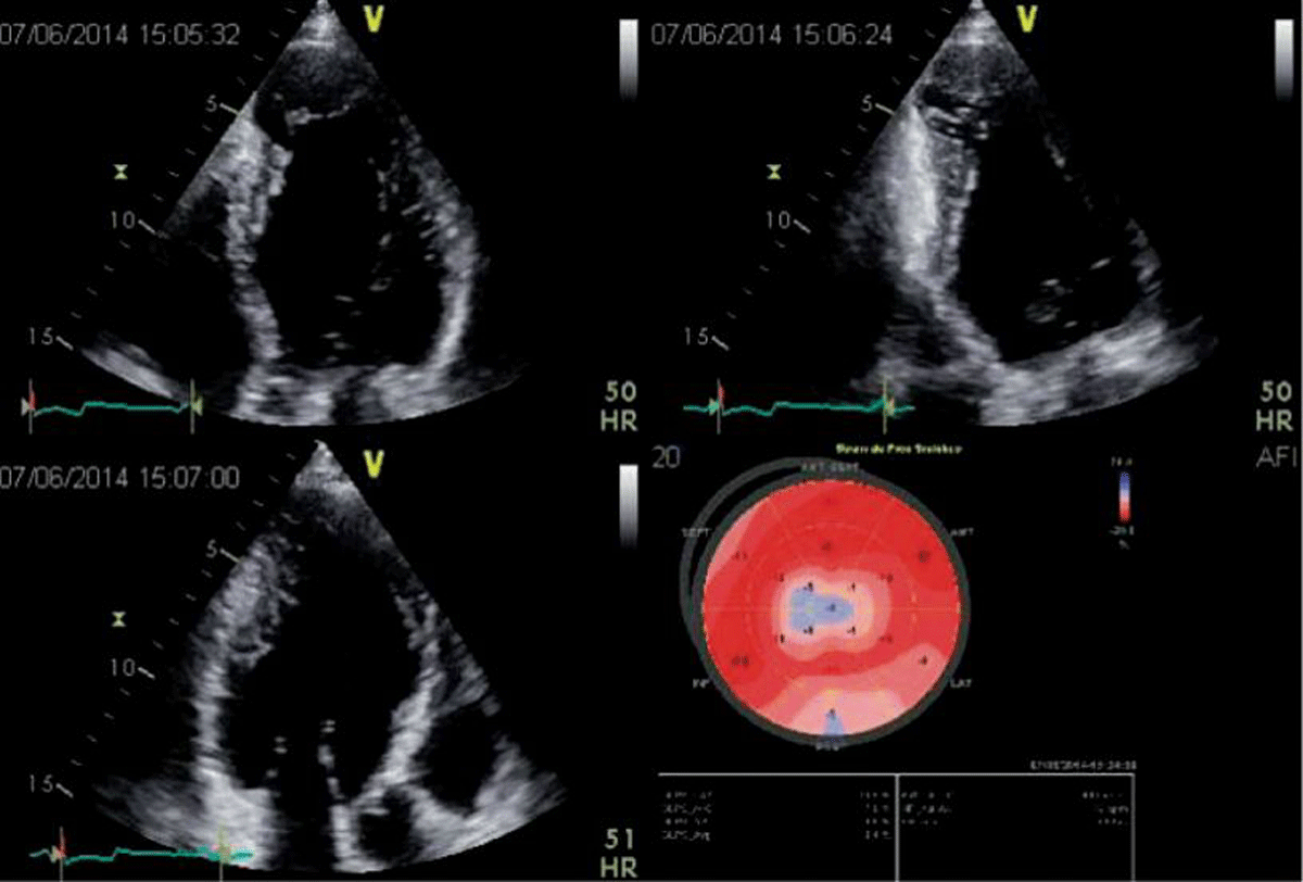
Figure 3
2D apical 4, 2 and 3-chamber views in a patient with Chagas disease and a typical aneurysm. In the 2-chamber view, a basal inferior aneurysm is also present. Longitudinal strain is abnormal in the apex, as well as on the basal inferior wall. Image: Marcia Barbosa. Reproduced with permission of the photographer.
Table 4
Recommendations for use of echocardiography in evaluation of left ventricle diastolic function.
| PARAMETER OR INDICATION | APPROPRIATE (A), INAPPROPRIATE (I) OR UNDETERMINED (U) |
|---|---|
| Evaluate diastolic function parameters in all patients with suspected ChD or ChCM | A |
| Index left atrial volume in all patients evaluated with color Doppler echocardiography | A |
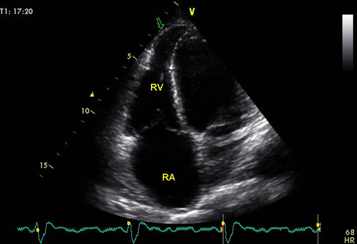
Figure 4
A young patient with Chagas disease presenting with biventricular dysfunction. Arrow in the 4 chamber-view, optimized to visualize the right ventricle, shows the infrequent finding of right ventricular aneurysm. Image: Marcia Barbosa. Reproduced with permission of the photographer.
Table 5
Recommendations for use of echocardiography in evaluation of the right ventricle.
| PARAMETER OR INDICATION | APPROPRIATE (A), INAPPROPRIATE (I) OR UNDETERMINED (U) |
|---|---|
| It is recommended to evaluate the dimensions and parameters of RV systolic function in all patients with Chagas disease when performing an echocardiogram. | A |
| Complement the evaluation of the right cavities with strain and 3D echo if these techniques are available | A |
| Estimate pulmonary systolic pressure in all patients with Chagas disease if possible | A |
| In the case of patients with Chagas disease and pacemakers or intracardiac devices, evaluate the functional repercussions at the level of the tricuspid valve and their adequate positioning. | A |
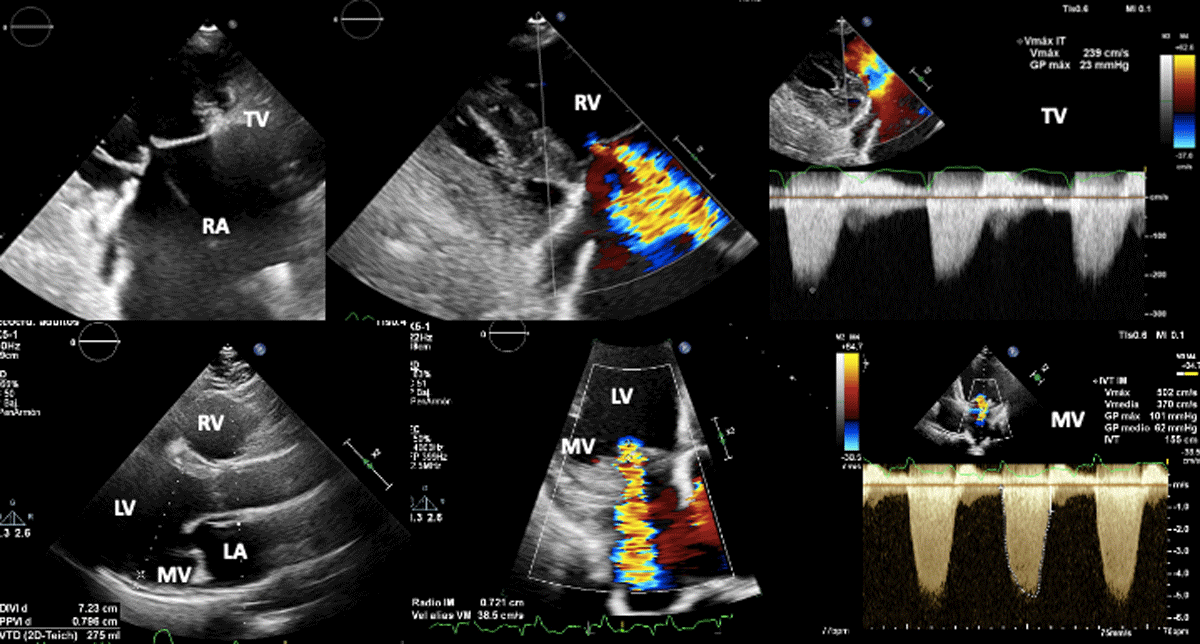
Figure 5
Biventricular dysfunction in a patient with Chagas disease. Superior figures show non coaptation of the tricuspid valve (due to right ventricular dilation) and its consequent functional regurgitation. Lower figures show mitral regurgitation secondary to left ventricular dysfunction.
Image: Mariana Corneli. Reproduced with permission of the photographer.
Table 6
Recommendations on when to perform an echocardiogram in patients with Chagas disease.
| SYMPTOMATIC STATE OF THE PATIENT | APPROPRIATE (A), INAPPROPRIATE (I) OR UNDETERMINED (U) |
|---|---|
| Asymptomatic patients with normal ECG should have a baseline echo, in a non-urgent fashion, where possible. | A |
| Asymptomatic patient with new changes in the ECG or patient that development symptoms that suggest cardiac compromise regardless of whether they have already undergone an echocardiogram previously | A |
| Asymptomatic patient without changes in the ECG with a previous echocardiogram performed less than 5 years ago without alterations | I |
| Repeat echocardiogram in patients with symptoms and impaired LV ejection fraction with impaired functional class | A |
| Repeat echocardiogram in symptomatic patients with LVEF <40% 3 to 6 months after adjusting medical treatment for heart failure to assess the impact of treatment | A |
| Repeat an echocardiogram annually in all patients with Chagas disease, regardless of the presence of electrocardiographic alterations or functional class, as an evolutionary control of the disease | I |