
Figure 1
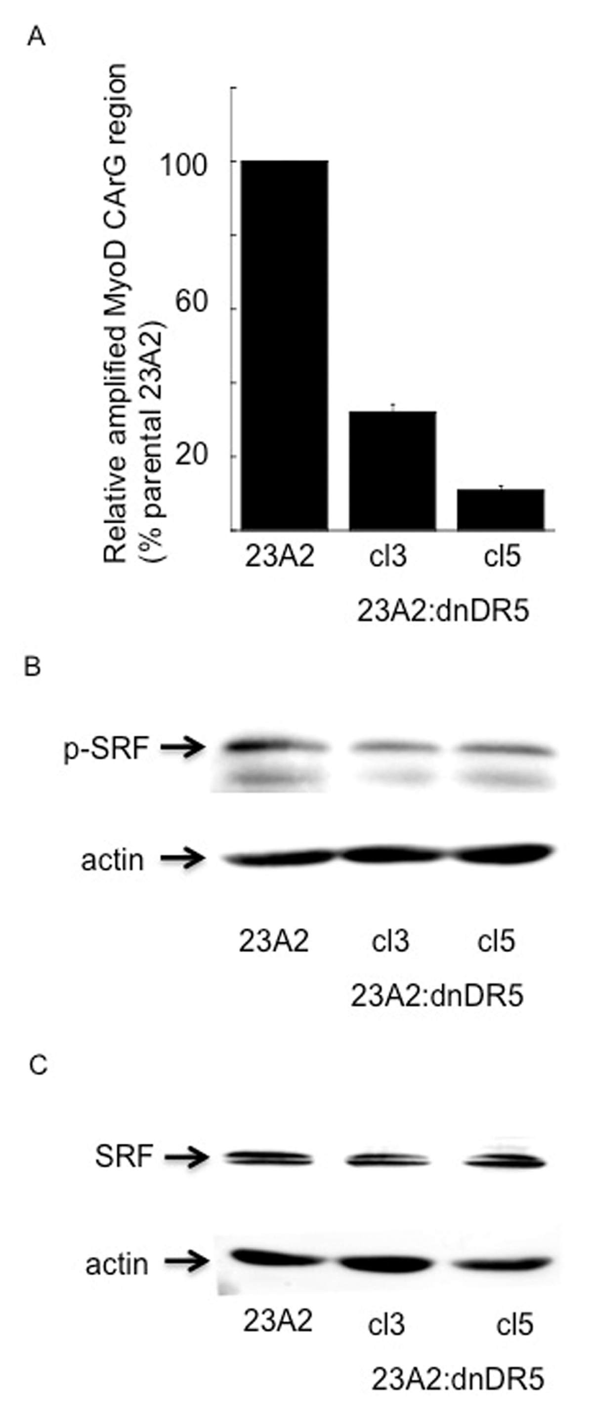
dnDR5 expression decreases SRF binding to the CArG box in the DRR of the MyoD enhancer as well as specific phosphorylation of SRF. In (A) chromatin from 1 × 10⁷ cells was cross-linked and digested with MNase to a length between 500–1000 bp. Chromatin Immunoprecipitation was performed on each cell sample using EZ ChIPTM Chromatin Immunoprecipitation Kit (Upstate) per manufacturer’s instructions. Chromatin from 2 × 10⁶ cells was immunoprecipitated as described in Methods. Quantitative PCR was used to assay for the relative levels of SRF binding near MyoD CArG element. Data was normalized to the signal detected from the input of each sample and presented as a percent of the signal obtained from parental 23A2 myoblasts. Error bars represent mean +/– SEM of triplicates. In (B) and (C), equal cell numbers were plated and the next day lysates were prepared and subjected to SDS-PAGE. Western analysis was performed using anti-phospho SRF, anti-SRF or anti-β-actin (loading and transfer control) and visualized as described in Methods. Shown are the results of one experiment that are representative of three independent experiments for (B) and two independent experiments for (C).
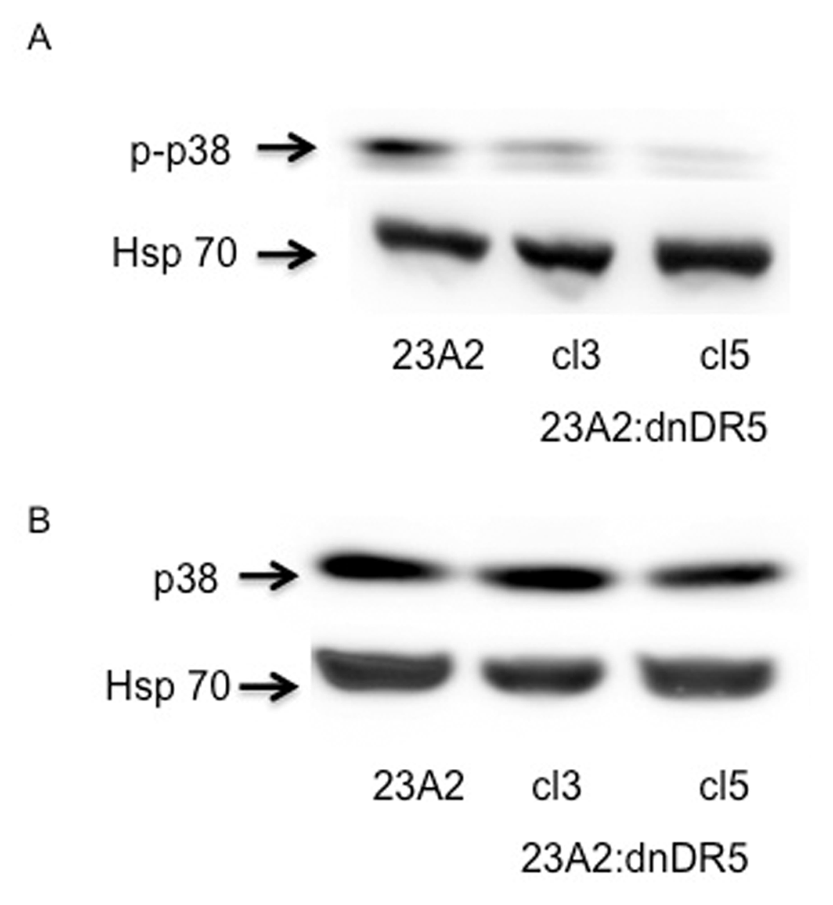
Figure 2
dnDR5 expressing myoblasts possess decreased levels of specifically phosphorylated p38. For each, equal cell numbers were plated and the next day lysates were prepared and subjected to SDS-PAGE. Western analysis was performed using anti-phospho p38 (A), anti-p38 (B), or anti-Hsp70 (loading and transfer control) and visualized as described in Methods. Shown are the results of one experiment that are representative of three independent experiments for (A) and two independent experiments for (B).
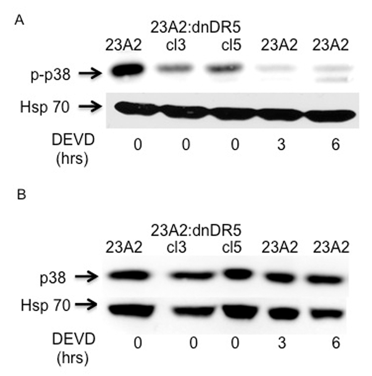
Figure 3
Myoblasts treated with a caspase 3 inhibitor possess decreased levels of specifically phosphorylated p38. For each, equal cell numbers were plated and the next day treated (or not) with DEVD-fmk as indicated in Methods. After the the indicated times, lysates were prepared and subjected to SDS-PAGE. Western analysis was performed anti-phospho p38 (A) or anti-p38 (B). Western analysis using anti-Hsp70 served as the loading and transfer controls. Bands were visualized as described in Methods. Shown are the results of one experiment that are representative of three independent experiments for (A) and two independent experiments for (B).
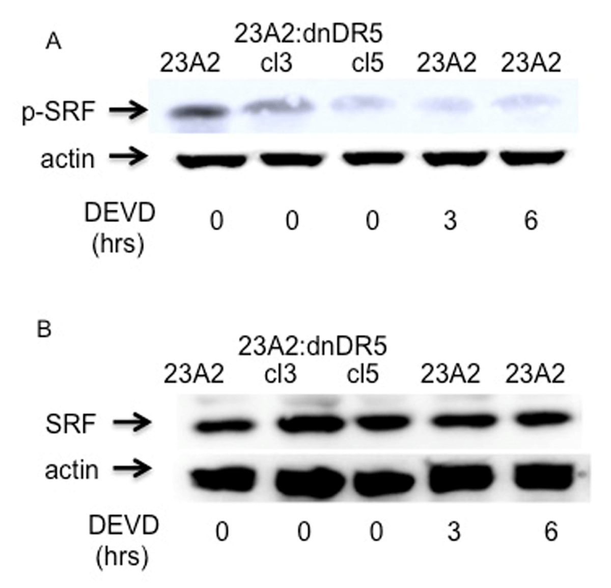
Figure 4
Myoblasts treated with a caspase 3 inhibitor possess decreased levels of specifically phosphorylated SRF. For each, equal cell numbers were plated and the next day treated (or not) with DEVD-fmk as indicated in Methods. After the the indicated times, lysates were prepared and subjected to SDS-PAGE. Western analysis was performed anti-phospho pSRF (A) or anti-SRF (B). Western analysis using anti-Hsp70 served as the loading and transfer controls. Bands were visualized as described in Methods. Shown are the results of one experiment that are representative of three independent experiments for (A) and two independent experiments for (B).
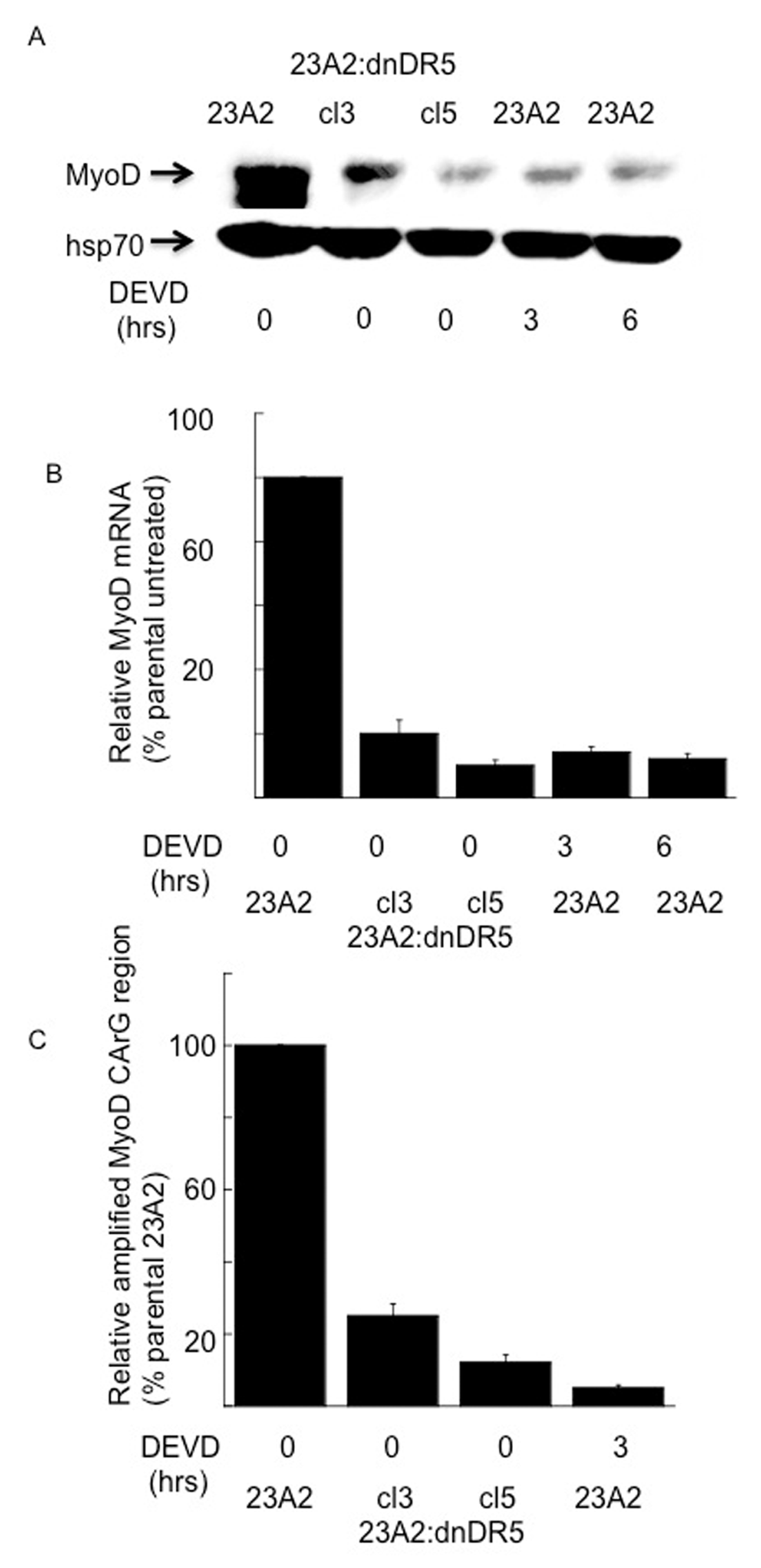
Figure 5
Myoblasts treated with a caspase 3 inhibitor possess decreased levels of MyoD protein, mRNA and SRF binding to the CArG box in the DRR of the MyoD enhancer. For each, equal cell numbers were plated and the next day treated (or not) with DEVD-fmk for the indicated times. In (A) Western analysis was performed using anti-MyoD or anti-Hsp70 (loading and transfer control) and visualized as described in Methods. Shown are the results of one experiment that are representative of three independent experiments. In (B) quantitative RT-PCR was used to assay for the relative levels of MyoD mRNA in total RNA samples derived from the indicated cell cultures. The Ct values for the MyoD PCR product were normalized to the Ct values for a β-actin product, run in parallel, as described in Methods. Error bars represent mean +/– SEM from triplicates. In (C), ChIP analysis was perfomed as described in the Figure 1 legend.
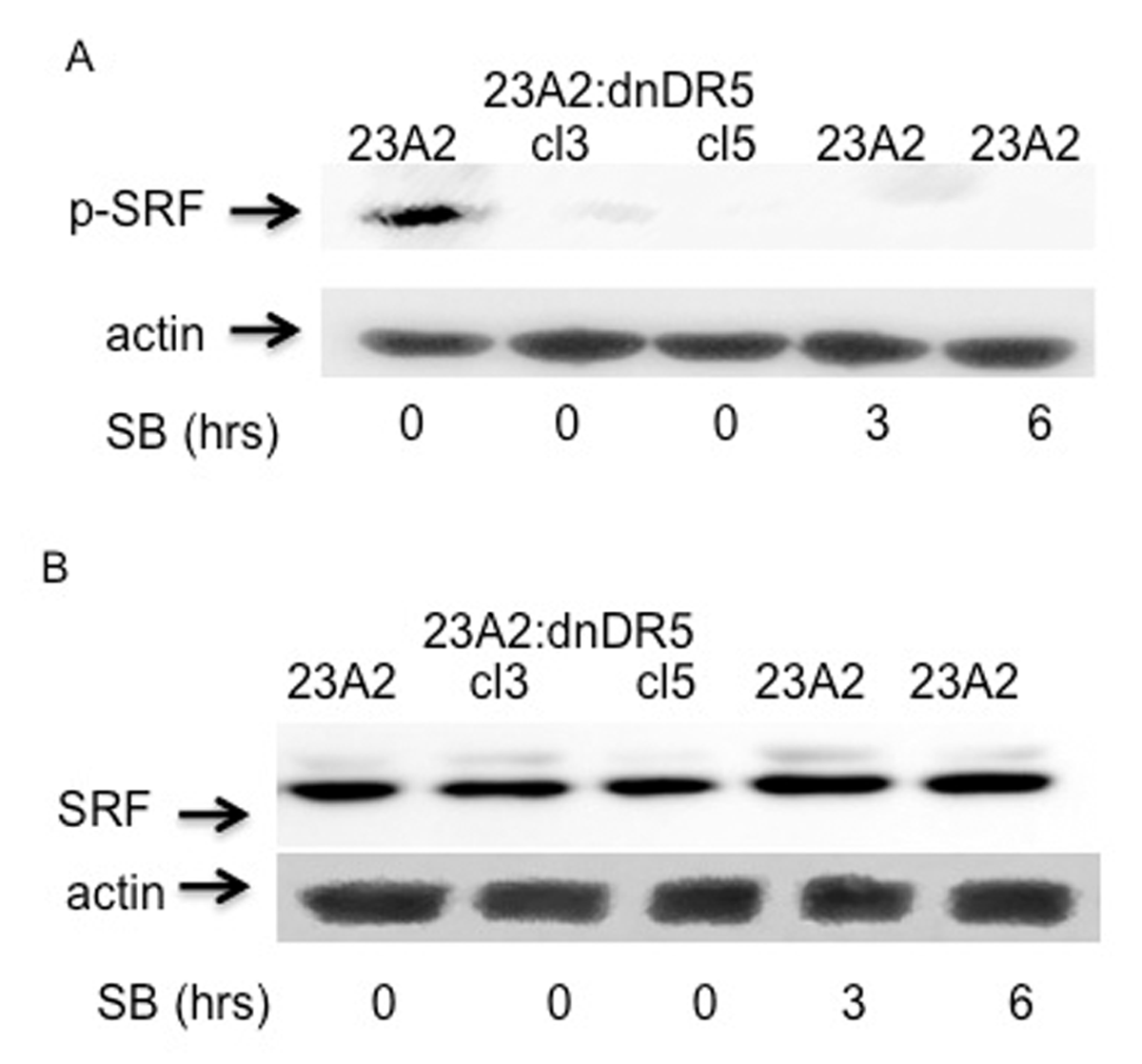
Figure 6
Myoblasts treated with a p38 inhibitor possess decreased levels of specifically phosphorylated SRF. For each, equal cell numbers were plated and the next day treated (or not) with SB 203580 for the indicated times. In (A) and (B), Western analysis was performed using anti-phospho SRF or anti-SRF, respectively with anti-actin serving as the loading and transfer control. Bands were visualized as described in Methods. Shown are the results of one experiment that are representative of three independent experiments for (A) and two independent experiments for (B).
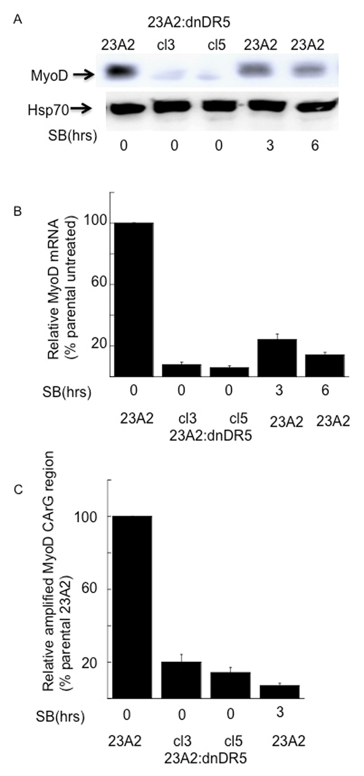
Figure 7
Myoblasts treated with a p38 inhibitor possess decreased levels of MyoD protein, mRNA and SRF binding to the CArG box in the DRR of the MyoD enhancer. For each, equal cell numbers were plated and the next day treated (or not) with SB 203580 for the indicated times. In (A), Western analysis was performed using anti-MyoD or anti-Hsp70 (loading and transfer control) and visualized as described in Methods. Shown are the results of one experiment that are representative of three independent experiments. In (B), quantitative RT-PCR was used to assay for the relative levels of MyoD mRNA as described in the legend to figure 5. In (C), ChIP analysis was perfomed as described in the Figure 1 legend.
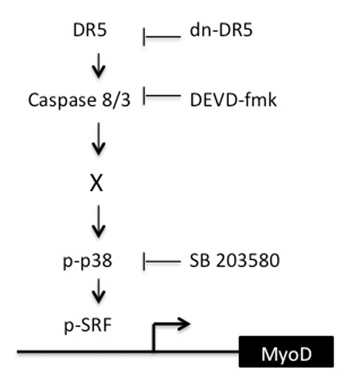
Figure 8
Model depicting the signalling pathway from DR5 to SRF-mediated transcription of MyoD. Basal signaling from DR5 to capases 8 and 3 results in specifically phosphorylated p38. Active p38 in turn leads to specifically phosphorylated SRF, SRF binding to the CArG box in the DRR of the MyoD enhancer, and expression of MyoD.