
Table 1
Primer sequences to generate dsRNAs.
| Primer Name | Sequence |
|---|---|
| Luc-F | taatacgactcactatagggAGATGGAACCGCTGGAGAGC |
| Luc-R | taatacgactcactatagggGACTCTGGCACAAAATCG |
| PERK-F | taatacgactcactatagggTGGCACAAGGAGGGGAAC |
| PERK-R | taatacgactcactatagggGCACCACTGGACCTAGTAAA |
| IRE1-F | taatacgactcactatagggCAAAAGCAGAGCGAGAATG |
| IRE1-R | taatacgactcactatagggTTAATGTCGCGATGCACAA |
| Atf6-I-F | taatacgactcactatagggAACATTACGGGATCAGATATTTC |
| Atf6-I-R | taatacgactcactatagggAAGGGTGGAGGTTCATTATATG |
| Atf6-II-F | taatacgactcactatagggCTTCAGCGTGTTCTGGTGAA |
| Atf6-II-R | taatacgactcactatagggGTTGGCAAAAGCCCTTTGTA |
| S1P-F | taatacgactcactatagggAAAGGTGTTGGAACTGTCGG |
| S1P-R | taatacgactcactatagggGGTATGCCGGAATAAGGGTT |
| S2P-F | taatacgactcactatagggGACAGAATTTTCGCGGAGAG |
| S2P-R | taatacgactcactatagggTGAGGTGCTACAATTCGCTG |
| TSC1-F | taatacgactcactatagggCAGCCTGCCAGAAATTAACT |
| TSC1-R | taatacgactcactatagggCCAGTCCTTCCACCGTCT |
Table 2
Primer sequences to quantitate total and spliced XBP1.
| Primer Name | Sequence |
|---|---|
| T-XBP1-F | ATACGCATCCTCGTCGAACATGGATGAC |
| T-XBP1-R | TCATCTAGAAAAACTCAGATCAAACTGG |
| S-XBP1-F | CCGAATTCAAGCAGCAACAGCA |
| S-XBP1-R | TAGTCTAGACAGAGGGCCACAATTTCCAG |
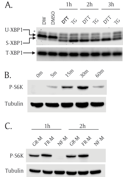
Figure 1
Both ER stress and TOR pathways are functional in S2R+ Drosophila cells. (A) ER stress inducers activated XBP1 mRNA splicing in S2R+ Drosophila cells. S2R+ cells were incubated with ER stress inducers for 1 hour, 2 hours, and 3 hours; then, total RNA was extracted and used to generate cDNA. Unspliced XBP1(U-XBP1) and spliced XBP1 (S-XBP1) were amplified using specific primer set across the XBP1 splicing region. Total XBP1 (T-XBP1) was used as a loading control. Asterisk (*) indicates a non-specific band. 1 mM dithiothreitol (DTT) and 0.2 μM thapsigargin (TG). Distilled water (DW) is the vehicle for DTT and Dimethylsulfoxide (DMSO) is the vehicle for TG. (B) Insulin increased phosphorylation of S6K. S2R+ cells were incubated with insulin (10 mg/ml) for multiple incubation times: 0 minutes, 5 minutes, 15 minutes, 30 minutes, and 60 minutes. The cell lysates were subjected to SDS-PAGE and transferred to nitrocellulose paper followed by Western blot analysis. Phospho(Thr398)-specific S6K antibody (P-S6K) detects only phosphorylated form of S6K. Tubulin serves as a loading control. (C) Nutrient-free (NF-M) media completely abolished S6K phosphorylation. S2R+ cells were cultured in growth media (GR-M) for 1 day. Then the cells were incubated with fresh media (FR-M) or nutrient free-media (NF-M) for 1 hour and 2 hours. Phospho(Thr398)-S6K and tubulin were detected by Western blot analysis.
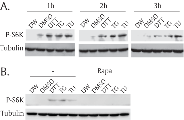
Figure 2
ER stress inducers activate the TOR pathway. (A) S2R+ cells were incubated in nutrient-free media for one hour, and then treated with three ER stress inducers in serum-free growth media for 1 hour, 2 hours, and 3 hours along with two vehicle controls. Then, cell lysates were prepared, subjected to SDS-PAGE, transferred to nitrocellulose paper, and then incubated with phospho(Thr398)-specific S6K (P-S6K) antibody or tubulin antibody. Distilled water (DW) was used as a vehicle for 1 mM dithiothreitol (DTT). The same amount of dimethylsulfoxide (DMSO) was used as a vehicle control for 0.2 μM thapsigargin (TG) and 0.5 mg/ml tunicamhycin (TU). (B) S2R+ cells were incubated in nutrient-free media for one hour and then, treated for another one hour with three ER stress inducers with or without 27 μM rapamycin (Rapa). Then, the cell lysates were subjected to Western blot.
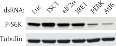
Figure 3
Knock down of Atf6 inhibits TOR signaling. S2R+ cells were transfected with luciferase (Luc), TSC1, eIF2a, IRE1, PERK, or Atf6 double-stranded (ds) RNA. The transfected cells were cultured for 2 days, and incubated with serum-free media for 1 hour before cells were collected. Then, Western blot analysis was performed with phospho(Thr398)-specific S6K antibody or tubulin antibody. Firefly luciferase (Luc), Tuberous sclerosis complex 1 (TSC1), Eukaryotic initiation factor 2a (eIF2a), Inositol requiring enzyme 1 (IRE1), Pancreatic ER kinase (PERK), and Activating transcription factor 6 (Atf6).
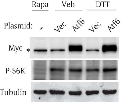
Figure 4
Overexpression of Atf6 activates the TOR pathway. S2R+ cells were transfected with an empty vector (Vec) or myc-Atf6 construct (Atf6); then, cultured for 2 days. The cells were incubated with vehicle or DTT in serum-free media for one hour. The cells were collected and subjected to Western blot. Rapamycin (Rapa) serves as a negative control for P-S6K. Asterisk (*) indicates a non-specific band in myc blot.
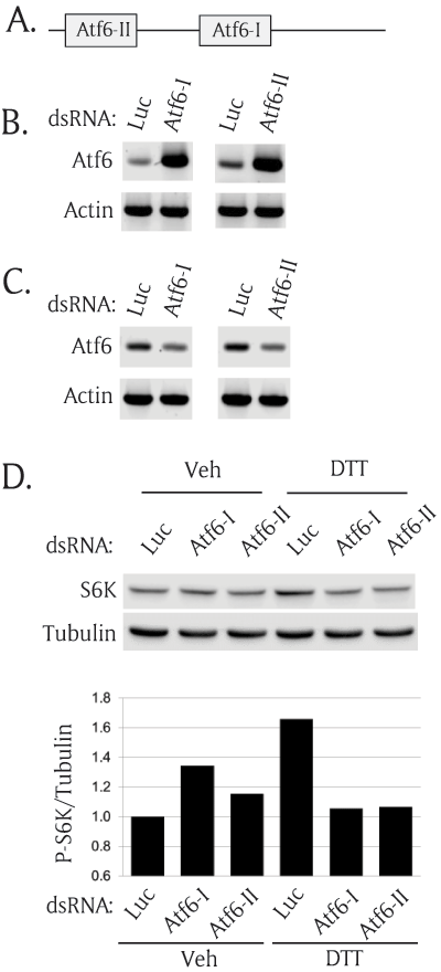
Figure 5
Atf6 is necessary for ER stress-mediated TOR activation. (A) A schematic diagram of two regions of Atf6 encoding double strand RNA (dsRNA) (B) Transfected Atf6 dsRNAs were present inside the cells 2 days after transfection. S2R+ cells were transfected with two regions of Atf6 dsRNAs and cultured for 2 days. The total RNAs were extracted and used to generate cDNA. Reverse transcriptase (RT)-PCR was performed to detect the transfected dsRNA by amplifying the inside sequence of the transfected dsRNA. Actin serves as endogenous control. (C) Atf6 dsRNAs knocked down endogenous Atf6 expression. To detect only Atf6 mRNA, the outside region of the transfected dsRNA was amplified using RT-PCR. Actin serves as an endogenous loading control. (D) Knock down of Atf6 abolished ER stress-induced phosphorylation of S6K. S2R+ cells were transfected with two different regions of Atf6 dsRNA. The transfected cells were cultured for 2 days and incubated with nutrient-free media for 1 hour. Then, the cells were incubated with vehicle or 1 mM DTT. The cells were collected and subjected to Western blot analysis. The Western blot results were quantitated and shown as the ratio of P-S6K/tubulin.
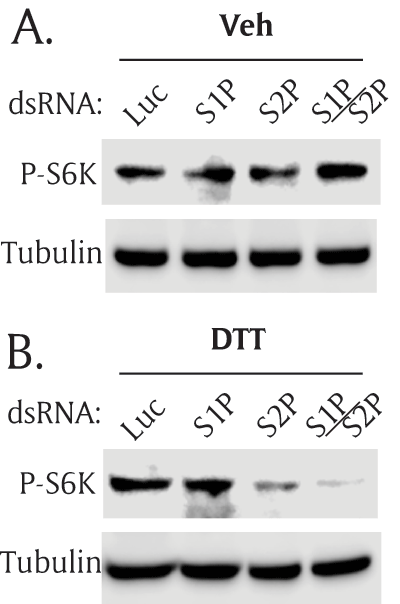
Figure 6
Both S1P and S2P proteases are necessary for ER stress-mediated TOR activation. S2R+ cells were transfected with double-strand (ds) RNA of S1P and S2P, separately or together. The transfected cells were cultured for 2 days, and treated with serum-free media (A) without ER stress, or (B) with ER stress. Then, Western blot analysis was performed with phospho(Thr398)-specific S6K antibody or tubulin antibody. Site-1 protease (S1P), Site-2 protease (S2P).