
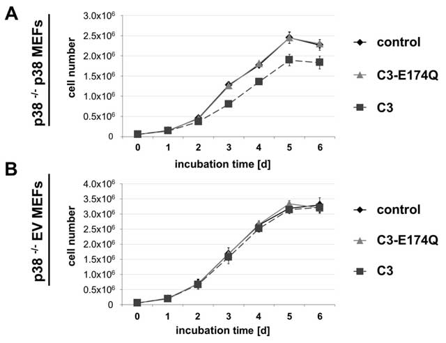
Figure 1
Influence of C3 on cell proliferation. For determination of the influence of C3 on cell proliferation, growth kinetic experiments were performed with both cell lines. p38 –/– p38 MEFs (A) or p38 –/– EV MEFs (B) were treated with 500 nM C3 or 500 nM C3-E174Q by replacing the medium including C3 or C3-E174Q every 48 h. The cell number was determined by trypan blue counting assay in duplicate after indicated incubation times. Growth curves represent mean values ± SEM of independent experiments (n = 3).
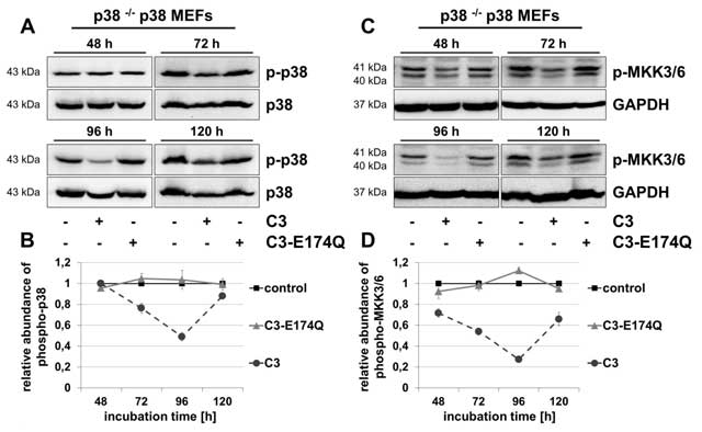
Figure 2
Reduced phosphorylation of p38 and MKK3/6 in C3-treated p38 –/– p38 MEFs. p38 –/– p38 MEFs were incubated with 500 nM C3 or 500 nM C3-E174Q for indicated time points, lysed and submitted to Western blot analyses for phospho-p38 (p-p38) and p38 (A, B), or phospho-MKK3/6 (p-MKK3/6) and GAPDH (C, D). (B) For densitometric quantification of p-p38, the signal intensity of p-p38 was adjusted to the corresponding intensity of p38. (D) The densitometric analysis of p-MKK3/6 was performed by normalizing the signal intensity of p-MKK3/6 of C3- and C3-E174Q-treated cells to the signal intensity of control cells. Representative Western blot analyses are illustrated. Results represent mean values ± SEM of independent experiments (n = 3).
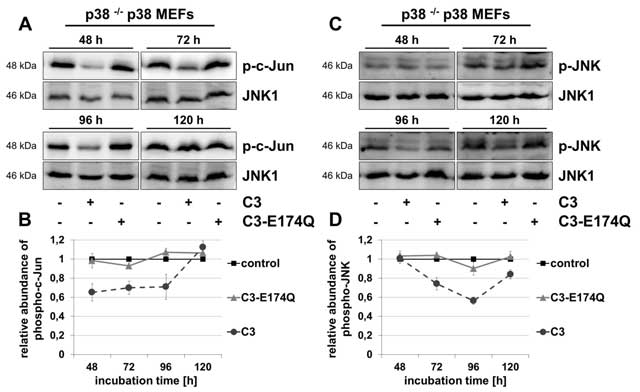
Figure 3
C3-induced decreased phosphorylation of c-Jun and JNK in p38 –/– p38 MEFs. p38 –/– p38 MEFs were incubated with 500 nM C3 or 500 nM C3-E174Q for indicated time points. Cells were lysed and submitted to Western blot analyses for phospho-c-Jun (p-c-Jun) (A, B), or phospho-c-Jun N-terminal kinase (p-JNK) and JNK1 (C, D). For densitometric analysis the signal intensity of p-c-Jun (B) or respectively p-JNK (D) was normalized to the corresponding intensity of JNK1. Representative blots are illustrated. Results depict mean values ± SEM of independent experiments of p-c-Jun (n = 3) and p-JNK (n = 4).
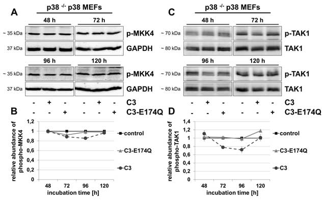
Figure 4
C3 marginally affected phospho-MKK4 and reduced the level of phospho-TAK1. After treatment of p38 –/– p38 MEFs with 500 nM C3 or 500 nM C3-E174Q for indicated incubation times, cells were lysed and applied to Western blot analyses for phospho-MKK4 (p-MKK4) and GAPDH (A, B), or phospho-TAK1 (p-TAK1) and TAK1 (C, D). (B) For densitometric quantification of p-MKK4 the signal intensity of p-MKK4 of C3- or C3-E174Q-treated cells were adjusted to the corresponding intensity of untreated control cells. (D) For quantification of p-TAK1 the signal intensity of p-TAK1 were normalized to the signal intensity of TAK1. Representing Western blot analyses are shown. Results illustrate mean values ± SEM of independent experiments (n = 3).
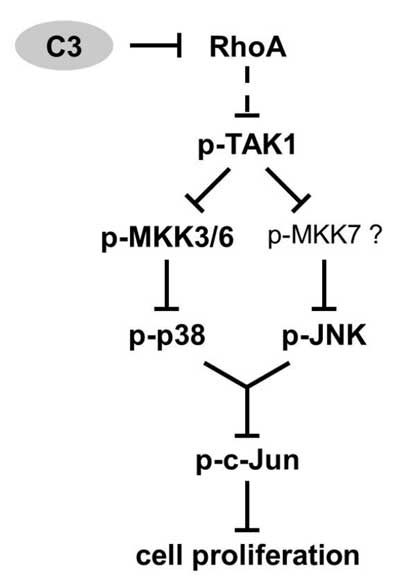
Figure 5
Proposed mechanism of C3-induced growth impairment. As a consequence of Rho inactivation by C3, the activity of MAPKKK TAK1 and downstream MKK3/6-p38 or respectively JNK pathway was decreased p38-dependently in p38 –/– p38 MEFs. Thus, the phosphorylation of c-Jun was reduced resulting in an impaired cell proliferation in C3-treated p38 –/– p38 MEFs.