
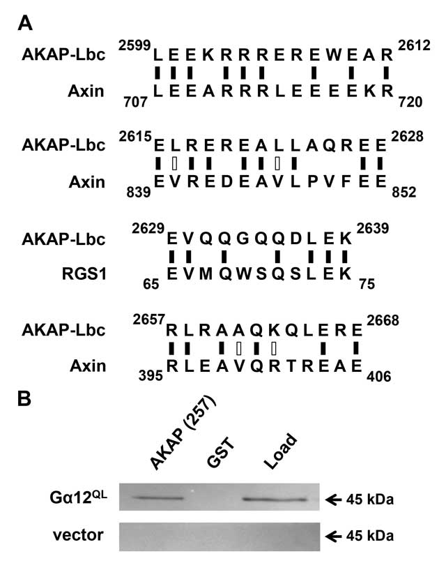
Figure 1
Identification of a Gα12-interacting region in AKAP-Lbc. (A) Sequence alignment of AKAP-Lbc with other Gα12 signaling targets. Results of Expasy SIM alignment using amino acid sequences of human proteins AKAP-Lbc (GenBank: NP_006729), axin-1 (NP_003493), and RGS1 (AAH15510) are shown. Regions of AKAP-Lbc excluding the tandem DH/PH domains were examined for homology to short sequences within RGS-1 and axin. Black vertical dashes indicate identical residues, open dashes indicate residues with similar properties. (B) Binding of Gα12 to an AKAP-Lbc region similar to axin and RGS1. HEK293 cells were transfected with a plasmid encoding myc-tagged, constitutively active Gα12 (GTPase-deficient Q229L mutant), or with pcDNA3.1 vector, and detergent-soluble extracts prepared as described in Methods. For each transfected sample, 5% of diluted extract was set aside as starting material (Load), and co-precipitation assays were performed using a Sepharose-bound GST-fusion of a 257-residue region of AKAP-Lbc, or GST alone. After SDS-PAGE and electroblot transfer, nitrocellulose membranes were probed with anti-Gα12 antibody (Santa Cruz Biotechnology; sc-409). Immunoblot images shown are representative of >5 experiments, with bands visible at the expected size (~45 kDa) for myc-tagged Gα12 and not detected in vector-transfected samples.
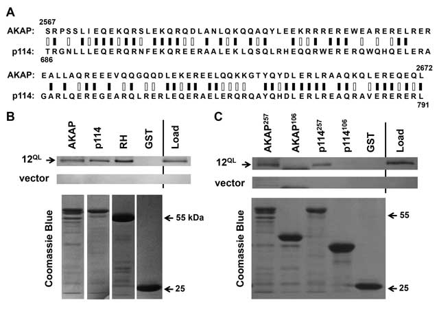
Figure 2
A conserved Gα12-binding domain in AKAP-Lbc and p114RhoGEF. (A) Sequence alignment of AKAP-Lbc and p114RhoGEF. Results of Protein BLAST (blastp) analysis are displayed, with black bars indicating identical residues (50/106), white bars indicating non-identical positive matches (28 additional residues), and zero gaps. (B) Interaction of Gα12 with regions of p114RhoGEF and AKAP-Lbc. Co-precipitation experiments were performed as described in Methods, using GST-fusions of the 257-amino acid C-terminus of AKAP-Lbc (AKAP), the corresponding 257 residues of p114RhoGEF (p114), amino acids 2-252 of p115RhoGEF containing its RH domain (RH), and GST alone. Load samples were set aside prior to addition of Sepharose-bound GST-fusion proteins. Co-precipitations from HEK293 cells transfected with myc-tagged, constitutively activated Gα12 (12QL) and empty vector were performed in parallel. For each sample, 20% of precipitated material was examined by SDS-PAGE/Coomassie Blue staining to assess levels of GST-fusion proteins, shown in lower panels. (C) Co-precipitations using the closely homologous, 106-amino acid domains within the Gα12-binding, 257-residue regions of AKAP-Lbc (AKAP) and p114RhoGEF (p114) are shown. Amino acid lengths of these adducts to GST are indicated as superscripts. Results in (B) and (C) are representative of three or more independent experiments.
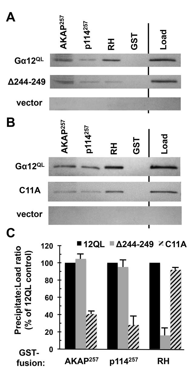
Figure 3
Gα12 binding to AKAP-Lbc and p114RhoGEF is distinct from RH-RhoGEF interaction. (A, B) Results of protein interaction experiments using mutant forms of myc-tagged, constitutively active Gα12 (Gα12QL). For both panels, Sepharose-immobilized AKAP-Lbc and p114RhoGEF constructs utilized in Figure 2B were tested, alongside the immobilized RH domain of LARG (RH), for ability to co-precipitate Gα12 variants from HEK293 cell extracts as described in Methods. Load samples were set aside prior to the precipitation step. Representative results are shown. (C) Immunoblot bands for Gα12 mutants were quantified using ImageJ, and for each sample the precipitate:load ratio was calculated and presented as a % of the precipitate:load ratio for non-mutated, constitutively active Gα12. Results shown are from three or more independent experiments, with graphs indicating mean ± standard error of the mean (s.e.m.).
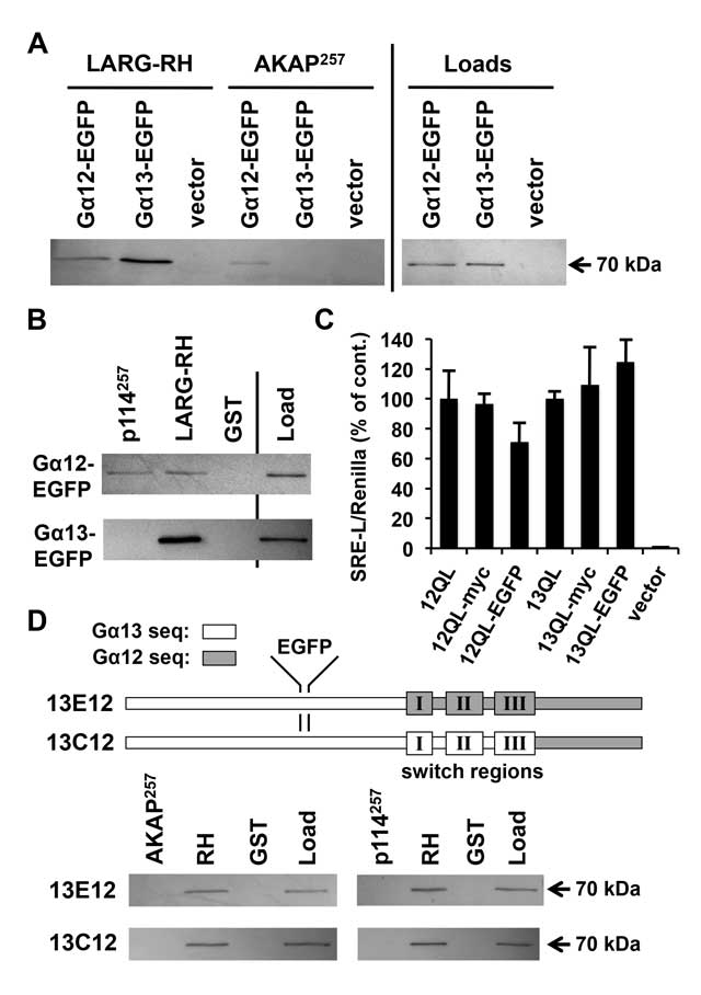
Figure 4
AKAP-Lbc and p114RhoGEF are selective for Gα12 binding. (A, B) Binding of EGFP-tagged G12/13 α subunits to RhoGEF domains. GST-fusions of the 257-amino acid AKAP-Lbc and p114RhoGEF regions used in Figure 2B, as well as the RH domain of LARG, were examined for ability to co-precipitate EGFP-tagged, constitutively active Gα12 and Gα13. Unmodified GST was tested as a negative control for α subunit co-precipitation. Shown are immunoblot analyses of precipitates and loads using an anti-EGFP antibody. All bands in these images co-migrated approximately with a 70 kDa protein standard (PageRuler; ThermoFisher). (C) Serum response element (SRE)-luciferase activation by epitope-tagged forms of constitutively active Gα12 and Gα13. Reporter constructs used were SRE-L, encoding firefly luciferase under control of a SRE-containing promoter, and pRL-TK, encoding Renilla luciferase governed by a thymidine kinase promoter. HEK293 cells grown in 12-well plates were transfected with SRE-L (0.2 µg) and pRL-TK (0.02 µg), plus plasmids encoding Gα12 or Gα13 as untagged, internally myc-tagged, or internally EGFP-tagged forms (0.1 µg). All constructs harbored activating Gln-to-Leu mutations in the switch II region, Q229L for Gα12 and Q226L for Gα13. Cells were harvested approximately 48 h post-transfection and assayed for luciferase activity as described in Methods. Firefly activity normalized for Renilla activity is shown for each sample (Y-axis) and presented as % of the untagged Gα12 or Gα13 response. Data presented are the mean of three independent experiments, with error bars indicating range. (D) Binding of Gα13/Gα12 chimeras to regions of RhoGEFs. A schematic of these chimeras, previously reported [47] and provided by Barry Kreutz (Univ. of Illinois at Chicago), is shown with an EGFP tag positioned as described in Methods. Both constructs harbor activating (QL) mutations in the switch II region. Lysates of HEK293 cells transfected with these tagged chimeras were subjected to co-precipitations by RhoGEF domains as described in (A) and (B), above. Immunoblot results using anti-EGFP antibody are shown, and are representative of three independent experiments.
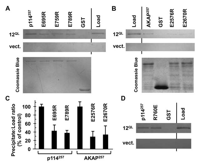
Figure 5
Conserved residues in AKAP-Lbc and p114RhoGEF participate in Gα12 binding. (A) Interaction of p114RhoGEF charge-reversal mutants with Gα12. The indicated mutants engineered in our GST-fused 257-amino acid region of p114RhoGEF were expressed in E. coli and immobilized on Sepharose. Concentrations of mutant p114RhoGEF proteins were adjusted so that approximately equal concentrations would be compared for ability to co-precipitate constitutively active Gα12 (12QL). For each sample, 20% of volume was analyzed by Coomassie Blue staining (lower image) to confirm uniform levels of immobilized p114RhoGEF variants. (B) AKAP-Lbc charge-reversal mutants were examined for Gα12 co-precipitation using the same procedure described for p114RhoGEF mutants. (C) For co-precipitations of Gα12, bands were quantified using ImageJ. Precipitate:load values for each mutant were calculated and presented as % of this value for non-mutated p114RhoGEF or AKAP-Lbc regions, which were set at 100%. Graphical data represent two or more independent experiments, with mean ± range shown. (D) Co-precipitation of Gα12 by an immobilized 257-residue p114RhoGEF construct harboring a charge-reversal of the Rgnef-homologous Arg residue (R760E). Images are representative of two independent experiments.
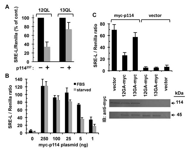
Figure 6
Preferential coupling of p114RhoGEF to Gα12 in signaling to SRE. (A) Effects of an ectopically expressed region of p114RhoGEF on Gα12- and Gα13-mediated signaling. HEK293 cells grown in 12-well plates were transfected with myc-tagged, constitutively active Gα12 or Gα13, plus either a construct encoding the Gα12-interacting 257 amino acids of p114RhoGEF with an N-terminal FLAG-tag (p114257) or empty vector. All cells were co-transfected with the firefly luciferase reporter SRE-L (0.2 µg) and the Renilla luciferase reporter pRL-TK (0.02 µg). Approximately 48 h post-transfection, cells were harvested and assayed for firefly and Renilla luciferase activity as described in Methods. For Gα12 and Gα13 transfections, effects of the p114RhoGEF construct are presented as % of vector control results, which were set at 100% for each α subunit. Graphs indicate mean ± s.e.m. for three independent experiments. (B) Effects of p114RhoGEF titration on serum dependence of its signaling to SRE. HEK293 cells grown in 12-well plates were transfected in duplicate with decreasing amounts of plasmid encoding myc-tagged, full-length p114RhoGEF (X-axis), plus uniform amounts of SRE-L and pRL-TK. After 32 h, one sample per transfection condition was washed twice with DMEM and serum-starved in the same medium for 12 h, whereas the duplicate sample received washes and addition of DMEM containing 10% FBS. Cell lysates were assayed for firefly and Renilla luciferase activity. Data shown are the mean of two independent experiments, with error bars indicating range. (C) Effects of dominant-negative Gα12 and Gα13 on serum-dependent signaling through p114RhoGEF. HEK293 cells were transfected with 5 ng plasmid encoding myc-tagged, full-length p114RhoGEF (myc-p114) or 5 ng empty vector, along with 50 ng plasmid encoding myc-tagged, constitutively GDP-bound variants of Gα12 (12GA-myc) or Gα13 (13GA-myc). All cells were co-transfected with SRE-L and pRL-TK and grown in DMEM + 10% serum for approximately 48 h, then assayed by luminometry. Data presented are mean ± s.e.m. for three independent experiments. At bottom are results of SDS-PAGE/immunoblot analysis of cell lysates, with expression of myc-p114RhoGEF (upper panel) and myc-tagged, dominant-negative G12/13 α subunits (lower panel) tracked using an anti-myc antibody. Immunoblots shown are a representative of three independent experiments.