
Figure 1
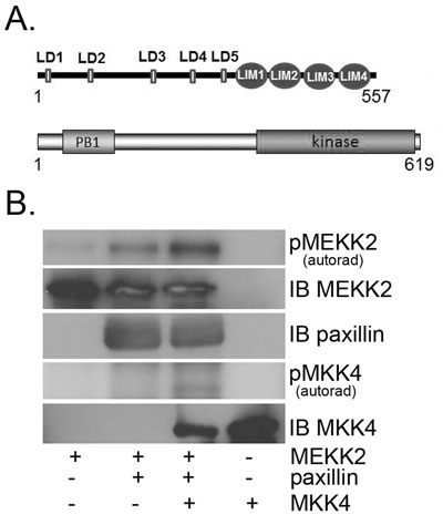
Paxillin promotes MEKK2 kinase auto-phosphorylation. A graphical representation of the domain structures of both MEKK2 and paxillin is shown in (A). (B) In vitro MEKK2 kinase assay analysis, with autoradiograph showing MEKK2 auto-phosphorylation (top panel), and MEKK2-dependent MKK4 phosphorylation (fourth panel). Anti-MEKK2 (second panel) and anti-paxillin (third panel) immunoblots were performed to demonstrate loading of reaction components. Results are representative of at least three independent experiments.
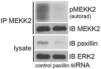
Figure 2
Paxillin expression knockdown inhibits MEKK2 activity. Displayed is an MEKK2 in vitro kinase assay with 32P ATP using MEKK2 auto-phosphorylation as an indication of kinase activity (top panel). MEKK2 was immunoprecipitated from 293T cells transfected with either paxillin siRNA or control siRNA. Anti-MEKK2 immunoblot shows the total amount of MEKK2 immunoprecipitated (second panel). Anti-paxillin immunoblot (third panel) shows siRNA-mediated expression knockdown, and anti-ERK2 blot demonstrates equal loading of lysate protein (bottom panel). Results are representative of at least three independent experiments.
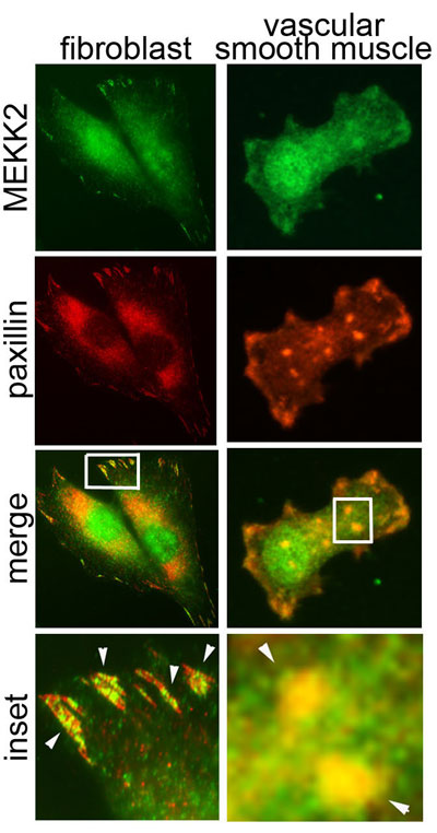
Figure 3
Fibronectin-induced co-localization of MEKK2 and paxillin in diverse cell types. Attachment to fibronectin induces MEKK2 to co-localize with paxillin in both murine fibroblasts (left column) and rat vascular smooth muscle cells (A7r5, right column). Cells were seeded on coverslips coated with fibronectin and allowed to attach for 6 hours, fixed and stained for immunofluorescence analysis with anti-MEKK2 (green) or anti-paxillin (red) antibodies. Arrowheads indicate areas of MEKK2/paxillin co-localization. Images are representative of at least three independent experiments.
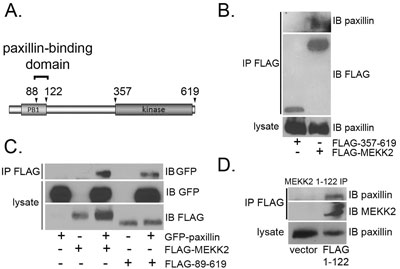
Figure 4
Mapping the paxillin binding site in MEKK2. (A) Diagram of MEKK2 protein with arrowheads indicating termini of truncation proteins used in these experiments. (B) Immunoblot analysis shows paxillin co-immunoprecipitating with FLAG-MEKK2 or FLAG-357-619 MEKK2. FLAG-tagged MEKK2 proteins were immunoprecipitated with anti-FLAG and co-immunoprecipitated paxillin was detected by anti-paxillin immunoblot (top panel). Endogenous paxillin present in each lysate was confirmed by anti-paxillin immunoblot (bottom panel), whereas the immunoprecipitated FLAG-tagged protein expression was detected by anti-FLAG blot (second panel). (C) FLAG-MEKK2 or FLAG-89-619 were co-transfected with GFP-paxillin, and the MEKK2 proteins were immunoprecipitated with anti-FLAG. Co-immunoprecipitated paxillin (top panel) was detected with anti-GFP blot. Expression of transfected proteins was confirmed with anti-FLAG and anti-GFP blots (second and bottom panels). (D) Cells were transfected with either empty vector or FLAG MEKK2 1-122, followed by immunoprecipitation of FLAG MEKK2 1-122 with anti-FLAG monoclonal antibodies. Co-immunoprecipitated paxillin from each lysate was detected by anti-paxillin immunoblot (top panel), as was total paxillin protein present in each lysate (bottom panel). Anti-FLAG immunoblot shows immunoprecipitated FLAG-1-122 (second panel). Results are representative of at least three independent experiments.
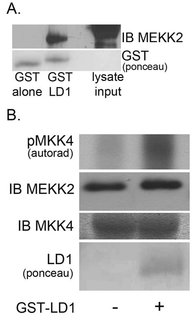
Figure 5
Paxillin LD1 motif peptide induces MEKK2 activity in vitro. LD1-GST pulldown of FLAG-MEKK2 from transfected 293T cell lysates is shown in (A) (upper panel) compared to an identical concentration of lysate pulled down with GST alone. Ponceau staining shows relative amounts of GST proteins used in the pulldown (lower panel). (B) MEKK2 in vitro kinase assay, with autoradiograph (top panel) showing rMKK4 phosphorylation +/- LD1-GST. FLAG-MEKK2 was immunoprecipitated from transfected cells with anti-FLAG antibodies and the activity assessed by in vitro kinase assays. Immunoblot analysis shows equal loading of MEKK2 (middle panel) and rMKK4 (bottom panel). Ponceau staining shows LD1-GST proteins included in the reaction (lower panel). Results are representative of at least three independent experiments.