
1. Introduction
Jump-landing is a common movement in sports and daily activities, yet it imposes substantial mechanical demands on the lower extremities. Upon ground contact, impact forces rise rapidly, and the lower limbs must absorb and dissipate the energy generated by body mass and jump height within a very short period. Peak vertical ground reaction force during landing can therefore reach several times body weight [1,2]. Effective impact attenuation depends primarily on coordinated flexion of the hip, knee, and ankle, as well as on the control of joint motion provided by eccentric muscle contractions during landing [3,4,5,6]. When this protective mechanism is insufficient, or when landing posture is poorly regulated, impact loads may be distributed less effectively across the lower-extremity joints and may increase the mechanical demand on passive structures around the ankle and knee. These landing-related mechanical characteristics are commonly considered relevant to lower-extremity loading and potential injury risk, including ankle sprains or instability and anterior cruciate ligament injuries [1,3,4].
Among the factors associated with landing mechanics, neuromuscular fatigue is considered particularly important. Previous studies have shown that fatigue can alter landing posture and movement-regulation strategies [7,8,9,10,11]. Under fatigued conditions, changes in sensory feedback, muscle response capacity, and force-generation ability may influence joint positioning, joint moment production, and lower-extremity loading patterns during landing [12,13]. Therefore, examining fatigue-related changes in landing kinematics and kinetics is important for understanding how individuals adjust lower-extremity loading when repeated jumping or landing tasks are performed under fatigue [9,11].
Although many studies support the influence of fatigue on landing biomechanics, the reported direction of these effects remains inconsistent. Some studies have reported greater ankle dorsiflexion together with increased hip and knee flexion after fatigue [7,14], whereas others have found greater ankle plantarflexion and reduced hip and knee flexion [8,10,15]. Findings on vertical impact loading are also inconsistent, with some studies reporting increased peak vertical ground reaction force after fatigue, whereas others have reported no significant change [5,16,17]. These discrepancies may be attributable to differences in fatigue-induction protocols, landing tasks, and participant characteristics across studies [9,16]. Importantly, they also suggest that the biomechanical effects of fatigue may vary across populations and may be influenced by participant-related factors such as age-related developmental stage.
Developmental differences may therefore be important when interpreting landing biomechanics under fatigue [1,18,19,20]. In jumping, landing, and rebound tasks, fatigue-related changes in landing biomechanics may not represent a response that is uniform across all age groups. From a physiological perspective, children and adults differ in strength production, neuromuscular function, sensorimotor development, and movement-regulation capacity [21,22,23,24]. These developmental characteristics may affect how the lower extremities coordinate joint motion and distribute mechanical loading during landing. In addition, recent child-adult fatigue studies have shown that children and adults differ in fatigability and in central and peripheral fatigue mechanisms [14,19,25]. Therefore, the way lower-extremity landing biomechanics are adjusted from pre- to post-fatigue may differ between children and adults. A direct comparison between these two age groups is not intended merely to describe age-group differences, but to determine whether developmental differences influence fatigue-induced changes in landing biomechanical regulation. Clarifying this issue may help distinguish whether fatigue-related landing changes represent a general fatigue effect across age groups or an age-specific regulatory pattern, thereby providing evidence for age-appropriate jump training, fatigue management, and landing-technique interventions.
Accordingly, the present study aimed to compare changes in lower-extremity landing biomechanics before and after jump-induced fatigue in children and adults under the same experimental conditions, in order to determine whether age-related developmental stage modifies the effects of fatigue on landing kinematics and kinetics. We hypothesized that jump-induced fatigue would alter lower-extremity landing biomechanics in both groups, but that children and adults would exhibit different fatigue-related landing adjustment strategies.
2. Materials and methods
2.1. Participants
An a priori power analysis was conducted using G*Power 3.1 for a repeated-measures analysis of variance examining the between-within interaction. The analysis was based on an effect size of f = 0.25, a significance level of α = 0.05, and a statistical power of 1 – β = 0.80. With two groups (children and adults) and two measurement conditions (pre-fatigue and post-fatigue), the minimum required sample size was 24 participants.
To allow direct comparison with previous jump-landing studies, school-aged children and young adults within age ranges comparable to those reported in previous studies were recruited [2,26]. The final sample comprised 24 participants, including 12 children (7 boys and 5 girls; age: 9.5 ± 1.5 years; height: 1.44 ± 0.08 m; body mass: 36.7 ± 6.7 kg; body mass index: 17.6 ± 2.0 kg/m2) and 12 adults (7 men and 5 women; age: 22.6 ± 0.5 years; height: 1.73 ± 0.04 m; body mass: 65.2 ± 7.3 kg; body mass index: 21.8 ± 1.6 kg/m2). All participants reported the right leg as their dominant leg.
Participants were eligible for inclusion if they had no history of lower-extremity musculoskeletal injury within the previous 6 months, had no relevant neurological disorder, and were able to complete the jump tasks independently. The study was approved by the Ethics Committee of Ningbo University (TY2025086). All adult participants and the parents or legal guardians of the children provided written informed consent after receiving a full explanation of the experimental procedures.
2.2. Experimental procedures
Before testing, participants completed 10 min of treadmill jogging at a self-selected speed, dynamic stretching, and supervised jump-task familiarization. They then wore a Polar H10 heart-rate strap (Polar H10, Finland) and rested quietly for 5 min; the final-minute mean heart rate was recorded as resting heart rate. Two experienced investigators attached 38 reflective markers (12.5 mm) according to the OpenSim Gait-2392 marker set.
The experiment included pre-fatigue testing, the fatigue intervention, and post-fatigue testing. During both testing stages, participants performed three successful bilateral countermovement jumps (CMJs). CMJ landing was selected as the assessment task because CMJ testing is widely used to evaluate lower-extremity neuromuscular function and fatigue-related changes [17,27,28], and it provides a standardized, self-initiated jump-landing movement that can be performed safely and consistently by both children and adults. Compared with drop-jump landing, CMJ landing also avoids potential confounding effects related to a fixed external drop height and box descent [29], thereby allowing fatigue-related changes in landing biomechanics to be assessed using the same pre- and post-fatigue task. For each trial, participants stood facing the force plates and performed a maximal-effort CMJ with a natural arm swing. Arm movement was not restricted to preserve a natural landing pattern, particularly in children. After landing, participants were required to stabilize their posture before stepping off the force plates. Trials were excluded if either foot missed the force plate or landed beyond its boundary.
Kinematic and kinetic data were collected using a 10-camera infrared Vicon motion-capture system (Oxford Metrics Ltd., Oxford, UK) and two Kistler force plates (model 9281B, Winterthur, Switzerland), sampled at 200 and 2,000 Hz, respectively. The fatigue protocol used between the two testing stages is described below.
2.3. Jump-induced fatigue protocol
A composite jump-fatigue protocol was used to reflect sport-related jumping demands and to impose repeated lower-limb loading under both slow and fast stretch-shortening cycle conditions [13,25,28]. CMJs and DJs were included because they are generally classified as slow (>250 ms) and fast (<250 ms) stretch-shortening cycle tasks, respectively [28,30]. Participants performed 20 consecutive CMJs followed immediately by 20 consecutive DJs from a 30-cm box, and these two sets constituted one fatigue cycle. Rest between cycles was strictly limited to 5 s, and participants were instructed to complete at least three cycles with maximal effort (Figure 1).
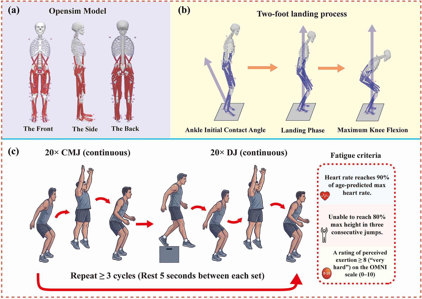
Figure 1
Overview of the musculoskeletal model and experimental procedures: (a) marker placement on the musculoskeletal model, (b) bilateral jump-landing task, and (c) fatigue protocol.
The fatigue protocol was terminated only when all three of the following criteria were met: (1) jump height fell below 80% of the participant’s maximal value for three consecutive jumps [11,29]; (2) heart rate reached at least 90% of the age-predicted maximal heart rate, calculated as 220 – age [18]; and (3) a rating of perceived exertion ≥ 8 (“very hard”) on the OMNI Rating of Perceived Exertion (OMNI-RPE) scale (0–10), which was used to reflect subjective fatigue during exercise [31]. For each participant, the number of completed cycles, total number of jumps, total duration of the fatigue protocol, final heart rate, final OMNI-RPE, percentage decline in jump height during the fatigue protocol, and the interval between fatigue termination and post-fatigue testing were recorded.
2.4. Data processing
Take-off and initial contact (IC) were identified as the instants at which the vertical ground reaction force fell below and subsequently exceeded 10 N [32], respectively. The landing phase was then defined as the interval from IC to the time of peak knee flexion.
Kinematic data were first preprocessed in Vicon Nexus (version 2.15.0 x64; Vicon Motion Systems, Oxford, UK). Custom Python scripts were then used to apply a fourth-order zero-lag Butterworth filter to the kinematic data with a cutoff frequency of 6 Hz. Ground reaction force data were low-pass filtered using a fourth-order zero-lag Butterworth filter with a cutoff frequency of 50 Hz. CMJ performance variables were calculated from the vertical ground reaction force data. Flight time was calculated as the interval between take-off and IC. Take-off velocity was derived from the vertical net impulse using the impulse-momentum method. Jump height was calculated from flight time using the following equation [17]:
Before inverse kinematics and inverse dynamics analyses, the generic Gait-2392 musculoskeletal model was individually scaled for each participant using the OpenSim Scale Tool based on a static calibration trial. Total model mass was adjusted to each participant’s measured body mass. The same marker set, scaling procedure, joint definitions, and coordinate system were applied to children and adults. Scaling quality was checked by visual inspection and marker-tracking errors during inverse kinematics; for all participants, the total root-mean-square marker error was below 2 cm and the maximum marker error was below 4 cm [33]. Inverse kinematics was performed to calculate lower-extremity joint angles, and inverse dynamics was conducted using the scaled model, inverse-kinematics results, and filtered ground reaction force data [33]. The resulting joint moments represented net internal joint moments. The kinematic variables of interest included joint angles in the subtalar frontal plane, ankle sagittal plane, hip sagittal and frontal planes, and knee sagittal plane. The subtalar frontal-plane variables represented subtalar inversion/eversion, with positive values indicating inversion and negative values indicating eversion. The kinetic variables of interest included vertical ground reaction force (vGRF), together with joint moments in the subtalar frontal plane, ankle sagittal plane, hip sagittal and frontal planes, and knee sagittal plane.For the discrete analyses, joint-angle values at initial contact, peak joint-angle values during landing, peak joint moments, and peak vertical ground reaction force were extracted. Peak vGRF was normalized to body weight (BW), whereas all joint moments were normalized to body mass (Nm/kg). All time-series data were normalized to 101 points across the landing phase to allow between-trial and between-subject comparisons.
2.5. Statistical analysis
Group differences in fatigue-protocol outcomes were examined using independent-samples t-tests. CMJ performance variables, including CMJ height, flight time, and take-off velocity, were analyzed using separate 2 × 2 mixed-design ANOVAs, with group treated as the between-subject factor and fatigue condition treated as the within-subject factor. For the biomechanical time-series data, a 2 × 2 mixed-design analysis of variance was performed within one-dimensional statistical parametric mapping (SPM1D), with group (children vs. adults) treated as the between-subject factor and fatigue condition (pre-fatigue vs. post-fatigue) treated as the within-subject repeated factor. Main effects of group and fatigue, together with the group × fatigue interaction, were examined. The SPM1D analysis was implemented using a two-factor ANOVA with one repeated measure (anova2onerm), with fatigue specified as the repeated factor. When necessary, within-group comparisons between pre-fatigue and post-fatigue conditions were performed using paired-sample SPM{t} tests, whereas between-group comparisons were performed separately under the pre-fatigue and post-fatigue conditions using two-sample SPM{t} tests.
For discrete (0D) variables, including peak vGRF and joint-angle values at initial contact and at peak, a separate 2 × 2 mixed-design ANOVA was used to examine main effects and the group × fatigue interaction. When a significant interaction was identified, simple-effects analyses with Bonferroni-adjusted post hoc comparisons were performed to control the type I error rate, and partial eta squared () was reported as the effect size for significant interaction effects. The significance level for all analyses was set at α = 0.05, and all statistical analyses were conducted in MATLAB (R2022a; The MathWorks, Natick, MA, USA).
3. Results
3.1. Fatigue-protocol outcomes
Fatigue-protocol outcomes are presented in Table 1. The time required to complete the fatigue protocol, number of completed cycles, and total number of jumps did not differ between children and adults. Final heart rate was higher in children than in adults, whereas final OMNI-RPE, the decline in jump height during the fatigue protocol, and the interval before post-fatigue testing showed no between-group differences. In both groups, final OMNI-RPE exceeded 8, the decline in jump height exceeded 20%, and post-fatigue testing began approximately 10 s after completion of the fatigue protocol.
Table 1
Fatigue-protocol outcomes in children and adults
| Variables | Child | Adult | P value |
|---|---|---|---|
| Completed cycles (N) | 3.42 ± 0.51 | 3.17 ± 0.39 | 0.192 |
| Total number of jumps (N) | 136.7 ± 20.6 | 126.7 ± 15.6 | 0.195 |
| Total duration of fatigue protocol (min) | 5.05 ± 0.65 | 4.86 ± 0.58 | 0.458 |
| Final heart rate (bpm) | 193.5 ± 5.2 | 184.6 ± 4.6 | < 0.001 |
| Final OMNI-RPE | 8.4 ± 0.5 | 8.6 ± 0.5 | 0.338 |
| Decline in jump height (%) | 23.8 ± 3.5 | 25.1 ± 3.8 | 0.393 |
| Interval before post-fatigue testing (s) | 10.6 ± 2.3 | 10.2 ± 2.0 | 0.654 |
Note. Values are mean ± standard deviation. OMNI-RPE, OMNI Rating of Perceived Exertion; post-fatigue testing interval, time from fatigue termination to post-fatigue testing.
3.2. Countermovement-jump performance
Countermovement-jump performance variables are presented in Table 2. CMJ height, flight time, and take-off velocity were higher in adults than in children. All three variables decreased after fatigue, indicating lower jump height, shorter flight time, and reduced take-off velocity in the post-fatigue condition. No significant group × fatigue interactions were observed for these variables.
Table 2
Countermovement-jump performance before and after fatigue in children and adults
| Variables | Child | Adult | P value | ||||
|---|---|---|---|---|---|---|---|
| Pre | Post | Pre | Post | Group | Fatigue | Interaction | |
| CMJ height (cm) | 24.41 ± 6.27 | 20.85 ± 5.70 | 38.47 ± 8.44 | 32.75 ± 7.25 | 0.002 | < 0.001 | 0.742 |
| Flight time (s) | 0.446 ± 0.058 | 0.412 ± 0.056 | 0.560 ± 0.061 | 0.517 ± 0.057 | < 0.001 | < 0.001 | 0.684 |
| Take-off velocity (m/s) | 2.18 ± 0.29 | 2.02 ± 0.28 | 2.74 ± 0.31 | 2.54 ± 0.29 | < 0.001 | < 0.001 | 0.706 |
Note. Values are mean ± standard deviation.
3.3. Landing-phase joint kinematics
Significant fatigue × group interactions were observed for the subtalar frontal-plane angle, ankle sagittal-plane angle, and hip frontal-plane angle during the landing phase. The interaction clusters occurred at 0.0–10.6% for the subtalar frontal-plane angle (F = 16.86, P = 0.047), 18.7–33.4% for the ankle sagittal-plane angle (F = 15.28, P = 0.024), and 15.9–59.4% for the hip frontal-plane angle (F = 19.15, P = 0.013). Within these clusters, adults showed greater subtalar inversion than children under the pre-fatigue condition; children exhibited greater ankle plantarflexion after fatigue, whereas adults showed greater dorsiflexion than children; and children showed greater hip adduction after fatigue and greater hip adduction than adults under the post-fatigue condition.
In addition, the hip sagittal-plane angle showed a significant main effect of fatigue at 4.9–46.0% of the landing phase (F = 22.52, P = 0.009), indicating reduced hip flexion after fatigue. The knee sagittal-plane angle showed significant main effects of group and fatigue, with clusters at 2.4–29.2% (F = 19.95, P = 0.006) and 0.1–39.6% (F = 36.34, P < 0.001), respectively, indicating greater knee flexion in adults than in children and reduced knee flexion after fatigue (Figure 2).
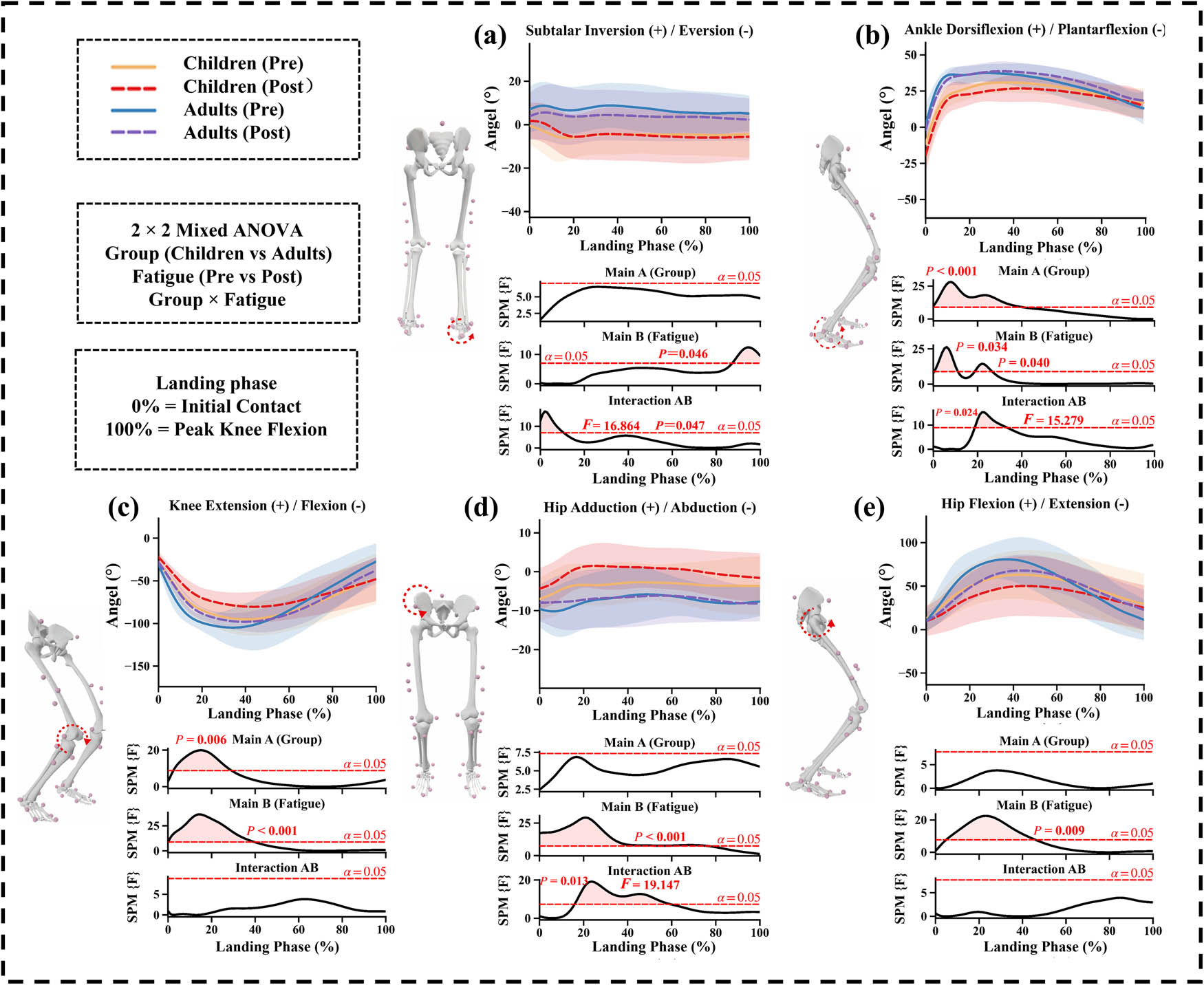
Figure 2
Landing-phase kinematic changes before and after jump-induced fatigue in children and adults: (a) subtalar frontal-plane angle, (b) ankle sagittal-plane angle, (c) knee sagittal-plane angle, (d) hip frontal-plane angle, and (e) hip sagittal-plane angle. Upper plots show mean ± standard deviation waveforms, and lower plots show SPM results; shaded regions indicate significant clusters.
3.4. Landing-phase joint kinetics
Significant fatigue × group interactions were observed for the subtalar frontal-plane moment and ankle sagittal-plane moment during the landing phase. The interaction clusters occurred at 0.6–20.5% for the subtalar frontal-plane moment (F = 18.78, P = 0.005) and 8.3–18.5% for the ankle sagittal-plane moment (F = 17.82, P = 0.016). Within the subtalar frontal-plane moment cluster, adults exhibited greater inversion moments after fatigue, whereas children exhibited greater inversion moments than adults only during the early pre-fatigue interval. Within the ankle sagittal-plane moment cluster, adults exhibited greater plantarflexion moments after fatigue, whereas children showed smaller plantarflexion moments than adults under the post-fatigue condition.
In addition, the knee sagittal-plane moment showed significant main effects of group and fatigue, with clusters at 2.0–27.8% (F = 31.48, P < 0.001) and 0.6%–8.1% (F = 16.61, P = 0.031), respectively, indicating greater knee sagittal-plane moments in adults than in children and reduced knee sagittal-plane moments after fatigue. The hip frontal-plane moment showed a significant main effect of fatigue at 6.9–18.4% of the landing phase (F = 15.64, P = 0.011), indicating reduced hip adduction moments after fatigue (Figure 3).
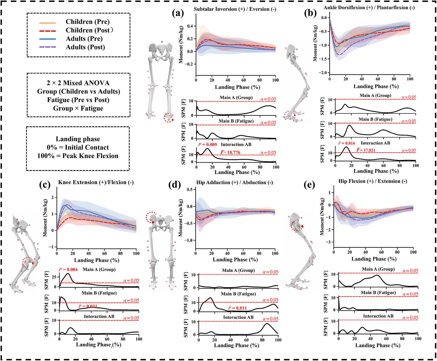
Figure 3
Landing-phase kinetic changes before and after jump-induced fatigue in children and adults: (a) subtalar frontal-plane moment, (b) ankle sagittal-plane moment, (c) knee sagittal-plane moment, (d) hip frontal-plane moment, and (e) hip sagittal-plane moment. Upper plots show mean ± standard deviation waveforms, lower plots show SPM results, and shaded regions indicate significant clusters.
3.5. Peak and initial contact joint angles
3.5.1. Peak joint angles
Significant fatigue × group interaction effects were observed for the peak ankle sagittal-plane angle and peak hip frontal-plane angle (F = 7.387, P = 0.013, ; F = 18.595, P < 0.001, ). For the peak ankle sagittal-plane angle, children showed a significant decrease after fatigue (Figure 4b; P = 0.003), whereas adults exhibited greater values than children under the post-fatigue condition (P < 0.001). For the peak hip frontal-plane angle, children showed a significant increase after fatigue (Figure 4e; P = 0.001) and greater values than adults under the post-fatigue condition (P = 0.007). Significant main effects of group and fatigue were observed for both the peak knee sagittal-plane angle (Figure 4c; F = 6.587, P = 0.018; F = 33.392, P < 0.001) and peak hip sagittal-plane angle (Figure 4d; F = 4.406, P = 0.048; F = 19.502, P < 0.001), indicating greater peak knee and hip flexion angles in adults than in children and lower values after fatigue.
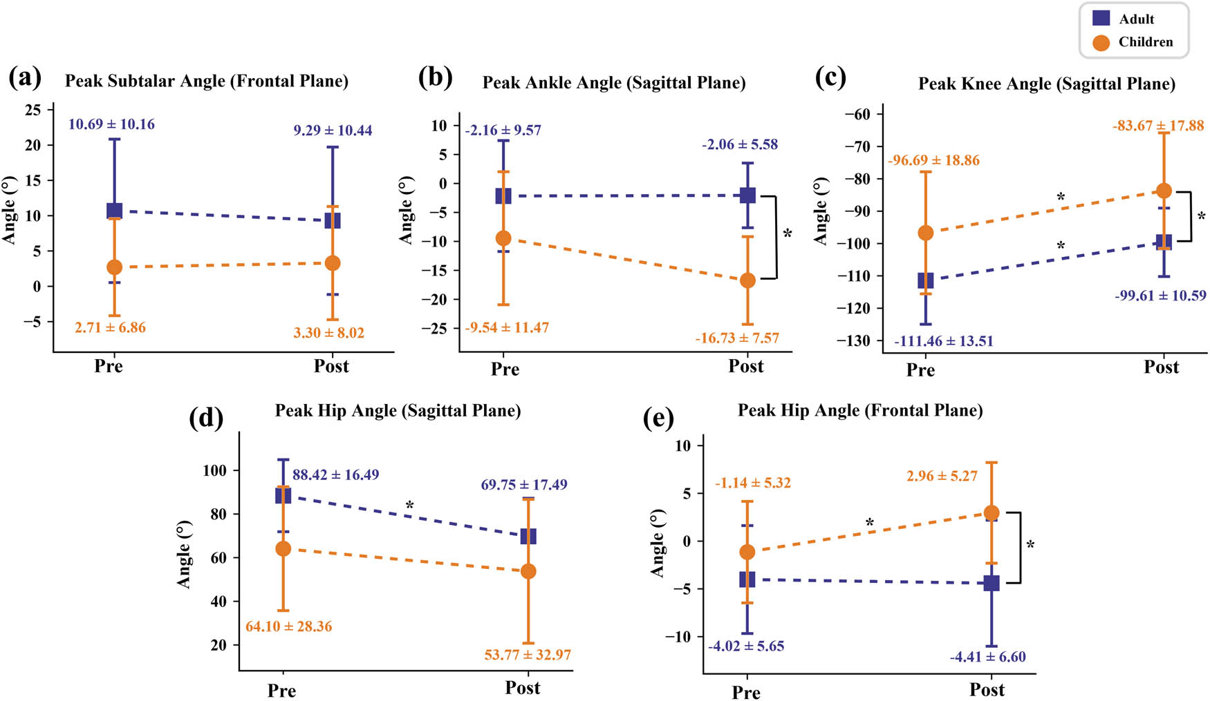
Figure 4
Peak joint angles before and after jump-induced fatigue in children and adults: (a) subtalar frontal-plane angle, (b) ankle sagittal-plane angle, (c) knee sagittal-plane angle, (d) hip sagittal-plane angle, and (e) hip frontal-plane angle. Values are mean ± standard deviation.
3.5.2. Initial contact joint angles
The ankle sagittal-plane IC angle (Figure 5b) showed significant main effects of group (F = 10.422, P = 0.004) and fatigue (F = 7.708, P = 0.011), indicating a more plantarflexed ankle posture in children than in adults overall and after fatigue compared with before fatigue. The knee sagittal-plane IC angle (Figure 5c) and hip frontal-plane IC angle (Figure 5e) showed significant main effects of fatigue only (F = 8.484, P = 0.008; F = 17.027, P < 0.001), with higher values after fatigue in both groups. No significant main effects were observed for the subtalar frontal-plane IC angle (Figure 5a) or hip sagittal-plane IC angle (Figure 5d).
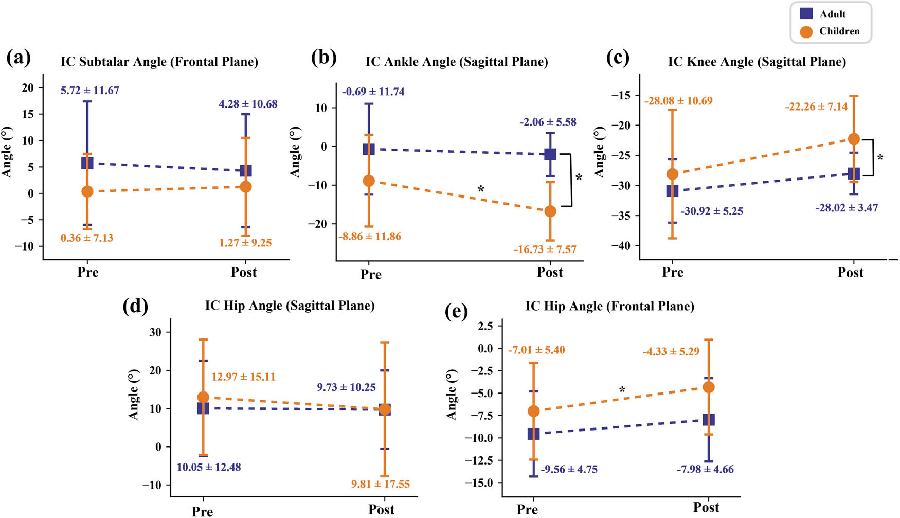
Figure 5
Initial-contact joint angles before and after jump-induced fatigue in children and adults: (a) subtalar frontal-plane angle, (b) ankle sagittal-plane angle, (c) knee sagittal-plane angle, (d) hip sagittal-plane angle, and (e) hip frontal-plane angle. Values are mean ± standard deviation.
3.6. Peak joint moments and vGRF
Significant fatigue × group interaction effects were observed for the peak subtalar frontal-plane moment and peak ankle sagittal-plane moment (F = 9.252, P = 0.006, = 0.296; F = 10.169, P = 0.004, = 0.316). After fatigue, children showed significant reductions in both peak subtalar frontal-plane moment (Figure 6a; P = 0.040) and peak ankle sagittal-plane moment (Figure 6b; P = 0.021). Under the post-fatigue condition, adults exhibited a greater peak ankle sagittal-plane moment than children (Figure 6b; P < 0.001). The peak knee sagittal-plane moment (Figure 6c) showed significant main effects of group (F = 25.708, P < 0.001) and fatigue (F = 11.147, P = 0.003), indicating greater values in adults than in children and lower values after fatigue. The peak hip frontal-plane moment (Figure 6e) showed a significant main effect of group only (F = 4.872, P = 0.038), with greater values in adults than in children.
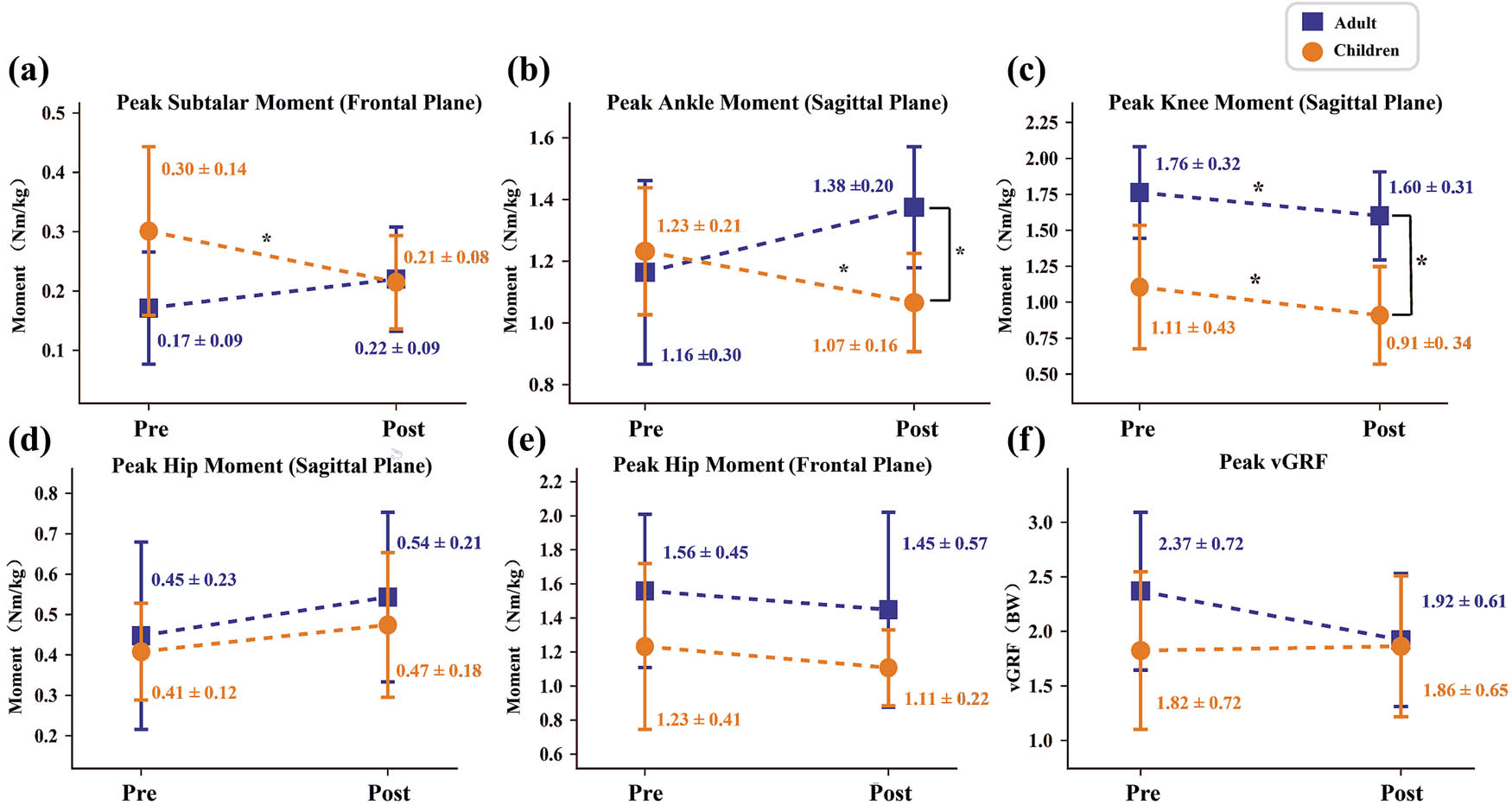
Figure 6
Peak kinetic variables before and after jump-induced fatigue in children and adults: (a) subtalar frontal-plane moment, (b) ankle sagittal-plane moment, (c) knee sagittal-plane moment, (d) hip sagittal-plane moment, (e) hip frontal-plane moment, and (f) vertical ground reaction force (vGRF). Values are mean ± standard deviation.
4. Discussion
The present study compared the effects of jump-induced fatigue on landing biomechanics in children and adults. Both groups reached the predefined fatigue endpoints, and the decline in CMJ performance confirmed the effectiveness of the fatigue protocol. Fatigue altered landing kinematics and kinetics in both groups, but the response differed by age. Children showed a more plantarflexed ankle posture, greater hip adduction, and reduced distal joint moments, particularly subtalar inversion and ankle plantarflexion moments. In contrast, adults retained greater hip and knee flexion and higher ankle plantarflexion moment output after fatigue. These findings suggest that children adopted a more constrained landing adjustment after fatigue, whereas adults preserved a more flexion-oriented strategy with greater ankle involvement. Overall, the results support the hypothesis that fatigue-induced changes in landing mechanics are age-dependent.
First, in the sagittal plane, both children and adults showed fatigue-related alterations in ankle angle, but the magnitude and direction of these changes differed between age groups. Children exhibited a more plantarflexed ankle posture after fatigue, as reflected by changes in peak ankle angle and significant time-series differences during early landing, whereas adults showed smaller changes in ankle sagittal-plane angle. Fatigue also reduced knee and hip flexion in both groups; however, adults still maintained greater hip and knee flexion than children. Taken together, these sagittal-plane results suggest that fatigue-related changes were mainly characterized by reduced hip and knee flexion, together with a more plantarflexed ankle posture in children. Because coordinated hip and knee flexion contributes to landing-load modulation and impact attenuation [1,34], these changes may indicate a more constrained sagittal-plane landing posture, particularly in children. In contrast, adults retained relatively greater hip and knee flexion, suggesting a more flexion-oriented landing adjustment after fatigue. This interpretation is consistent with Swartz et al., who reported that adults typically land with greater knee and hip flexion than children [2], and with Toussaint et al., who suggested that younger children show greater early-landing stiffness and rely more on passive tissues for energy absorption [35].
The changes in lower-extremity joint moments support these findings. After fatigue, children showed a significant reduction in peak subtalar frontal-plane moment and a slight decrease in peak ankle plantarflexion moment. The reductions in subtalar frontal-plane moment and ankle plantarflexion moment may together suggest altered distal joint loading in children under fatigued conditions, which may be related to ankle posture regulation and landing-load modulation during landing. In contrast, adults demonstrated greater subtalar frontal-plane and ankle plantarflexion moments after fatigue. This finding is consistent with the results of Orishimo and Kremenic, who reported that knee joint moments decreased after fatigue, whereas ankle joint moments increased correspondingly [36]. This pattern may suggest a distal compensatory strategy, whereby adults showed greater ankle joint involvement in regulating landing loads under fatigued conditions.
Hip moment changes were limited, with a fatigue effect observed only in the frontal plane. By contrast, the knee sagittal-plane moment showed a significant fatigue effect. After fatigue, both children and adults exhibited a reduction in knee sagittal-plane moment, although adults still showed higher knee sagittal-plane moment values than children overall. In the present study, children showed a decrease in knee sagittal-plane moment together with reduced knee flexion after fatigue. This finding is consistent with Briem et al., who reported reduced knee flexion excursion and lower knee flexion moments during post-fatigue drop-jump landings in children [26]. Taken together, these findings may suggest that fatigue reduced the sagittal-plane knee motion available for landing-load modulation in children [14,26], contributing to a more constrained sagittal-plane landing posture. This interpretation is also in line with previous work showing that fatigue may reduce knee flexion during landing in children, whereas adults may increase knee flexion as part of a more attenuating landing strategy [14]. However, because vertical ground reaction force did not change significantly in the present study and showed only a slight increasing trend, this pattern should be interpreted as a cautious indication of a relatively harder landing tendency rather than direct evidence of increased impact loading. In contrast, although adults also showed a reduction in knee sagittal-plane moment, their absolute values remained higher than those of children, and they still maintained relatively greater knee and hip flexion after fatigue. Together with the increased ankle moment response discussed above, adults may have adopted a landing adjustment strategy characterized by greater hip and knee flexion and greater ankle joint involvement under fatigued conditions.
Although this study provides novel insights, several limitations should be acknowledged. First, the standardized jump-landing task was performed in a laboratory setting and may not fully represent real sport situations. Second, the analysis was limited to kinematic and kinetic characteristics, with only sagittal-plane knee variables examined and no simultaneous assessment of muscle activity. Therefore, the underlying neuromuscular mechanisms should be interpreted cautiously. Future studies should use more sport-specific tasks and combine multiplanar kinematics with electromyography to further clarify age-related differences in fatigued landing.
5. Conclusion
Jump-induced fatigue altered landing kinematics and kinetics in both children and adults, but the response differed between age groups. After fatigue, children showed a more constrained landing adjustment, characterized by greater ankle plantarflexion, greater hip adduction, reduced hip and knee flexion, and lower subtalar and ankle moments. In contrast, adults retained greater hip and knee flexion and higher ankle moment output, suggesting a more flexion-preserving landing strategy. These findings indicate that age-related developmental differences influence landing regulation under fatigue and should be considered in age-specific jump training and injury-prevention programs.
Acknowledgments
The authors thank all participants involved in this study.
Funding information
This study was sponsored by National Key R&D Program of China (2024YFC3607305), General Program of Guangdong Provincial Natural Science Foundation of China (2026A1515010084), Joint Fund of Zhejiang Provincial Natural Science Foundation of China (LKLY26H170001), Zhejiang Provincial Key Project of Education Science Planning (2025SB084), Scientific Research Fund of Zhejiang Provincial Education Department (Y202559510), Ningbo Key Research and Development Program (2022Z196), Zhejiang Rehabilitation Medical Association Scientific Research Special Fund (ZKKY2023001), Research Academy of Medicine Combining Sports, Ningbo (No.2023001), Ningbo Clinical Research Center for Orthopedics and Exercise Rehabilitation (No.2024L004), Start-up Research Funding from Ningbo University (ZX2026000474), and K. C. Wong Magna Fund in Ningbo University.
Author contributions
A. The preparation of the research program: Xiaofeng Lin, Zifan Xia, Yihan Qian, Dong Sun, Yang Song, Yaodong Gu; B. The execution of research: Xiaofeng Lin, Zifan Xia, Yihan Qian, Xuanzhen Cen, Dong Sun; C. The statistical analysis: Xiaofeng Lin, Zifan Xia, Yihan Qian, Shunhang Wang, Yufan Xu, Xuanzhen Cen, Danica Janicijevic, Yang Song, Dong Sun, Yaodong Gu; D. The interpretation of data: Xiaofeng Lin, Zifan Xia, Yihan Qian, Shunhang Wang, Yufan Xu, Xuanzhen Cen, Danica Janicijevic, Yang Song, Dong Sun, Yaodong Gu; E. Preparation of the manuscript: Xiaofeng Lin, Zifan Xia, Yihan Qian, Shunhang Wang, Yufan Xu, Danica Janicijevic, Yang Song, Yaodong Gu; F. Obtain financing: Yang Song, Yaodong Gu.
Conflict of interest statement
Authors state no conflict of interest.
Data availability statement
The data supporting the findings of this study are available from the corresponding author upon reasonable request.