
1. Introduction
The masticatory system serves as a unique work tool for wind instrumentalists. Therefore, its condition – both functional and structural – affects not only physical well-being but also quality of life, directly impacting professional ability and daily functioning. During years of playing the instrument, saxophonists engage in repetitive motor activity, which can be considered monotypic work. The similarity of the cycles refers to the temporal sequences, developed strength, muscle tension, and spatial characteristics of the movements. A work cycle corresponds to a motor pattern repeated when playing specific pieces or performing exercises using the same instrument in the same (standing) position. Both the length of the cycle and the number of phases within it influence the load on the musculoskeletal system [1,2]. In a review of the literature by Woźniak and Piątkowska [3], it is indicated that the most frequently studied muscles of the masticatory system are the masticatory and temporal muscles. This also finds support in the work by Castroflorio et al. [4] and in a review of Nęcka’s [5] publications on muscle activity in the stomatognathic system regarding electromyographic studies. According to Woźniak and Piątkowska [3], the temporal muscles show the greatest activity in the resting position of the mandible, while the masticatory muscles are most active when the mandible performs adduction movements, which are necessary for holding the saxophone mouthpiece after opening the mouth. Therefore, it seems reasonable to examine the tension of the masseter muscles during saxophone-playing activities. These muscles are responsible for pressing the mandible against the maxilla and enhancing bite force, which, in this case, translates into stabilisation of the mouthpiece. Tecco et al. [6] examined the facial and sternocleidomastoid muscles, as well as the trapezius muscle, playing postural and functional roles by stabilising the shoulder joint and elevating the arm [7,8]. Klich et al. [9] assessed the tension of the descending part of the trapezius muscle during repetitive activities requiring the elevation of the upper limbs. Holding the instrument during repeated, daily, multi-hour saxophone practice can be considered such an activity. Interviews with the musicians enrolled in this study indicated that the upper portion of the trapezius muscle was the most tense and painful area, especially after practice and concerts, which were also subject to stress. These considerations formed the basis for selecting the masseter muscles and the descending portion of the trapezius muscle for surface electromyographic analysis. The authors assumed that knowledge on muscle tensions accompanying activities related to playing the saxophone is necessary both from the perspective of effectively managing the effort of the musculoskeletal structures involved in playing and in the context of professional overload prevention. Therefore, the aim of this study was to investigate whether saxophonists experience muscle tone variations in the masseter muscles and the descending parts of the trapezius dorsi muscle in resting conditions and during activities that load the stomatognathic system. This work could provide a basis for filling the existing research gap. The results presented herein may be of significance to both science and physiotherapeutic practice, supporting the development of programmes for the prevention of postural defects in musicians playing wind instruments.
2. Material and methods
2.1. Participants
The 2024 study included 30 men aged 20–30 years (average age: x = 25.27 ± 2.88 years), students of saxophone classes at the Jazz and Popular Music Department of randomly selected Music Academies in Poland (average saxophone playing experience x = 12.57 ± 2.06 years).
Before commencing the study, the following inclusion criteria were established: male sex; age between 20 and 30; saxophone playing experience of 10–15 years; playing the saxophone in a standing position 7 days a week, at least 5 hours per day; right-handedness and right-footedness; not playing the saxophone within 10 hours preceding the study; and providing written, informed consent for participation in the study. Men with neuromuscular or other chronic diseases affecting the musculoskeletal system, cognitive impairments or dementia, or congenital skeletal defects, were excluded from the study.
2.2. Procedures
Tension of selected masticatory and dorsal muscles, at rest and during functional loading of the stomatognathic system, was measured using the Noraxon Ultium EMG (Noraxon, Inc., Scottsdale, Arizona, USA). sEMG data were processed using MyoResearch XP software for recording and analysing data from Noraxon surface electromyography systems. The license to perform the research was provided by Skyfi sp. z o.o (Gliwice, Poland). The intraclass correlation coefficient for measurements using this device is ICC = 0.85–0.97 [10]. The 4-channel version of the Noraxon Ultium EMG electromyograph enabled the simultaneous diagnosis of two muscle groups. Action potentials were recorded from the skin surface for the masseters and the descending part of the trapezius muscle. The electrode orientation was parallel to the muscle fibres. Active electrodes were placed as shown in Figure 1, taking anthropometric points located in the central part of the muscle bellies into account. These points were identified by palpation in the area of greatest muscle tension during isometric contraction [11].
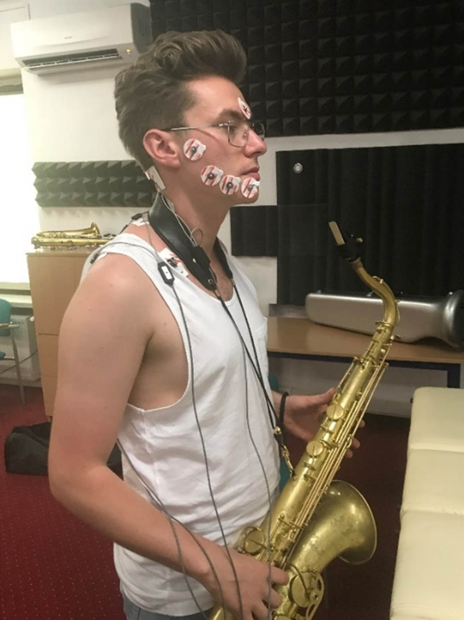
Figure 1
Participant during surface electromyography recording (Source: the authors’ archive; the participant provided written informed consent for publication of the photograph).
For the masseters, isometric contraction was achieved by clenching the teeth, and to determine the electrode placement on the muscle, palpation was performed 3 cm superiorly, anterosuperiorly, from the angle of the mandible, parallel to an imaginary line connecting the inferior angle of the jaw with the outer corner of the eye on the same side of the face [11]. For the descending part of the trapezius muscle, to achieve an isometric contraction, the subject performed an ipsilateral shoulder raise along with lateral flexion and contralateral cervical rotation against resistance to the shoulder and head applied by the examiner, in the direction opposite to the subject’s movements. To determine the electrode placement on the trapezius dorsi, palpation was performed midway between the seventh cervical vertebra and the acromion, along the course of the muscle fibres [12].
Electrodes from channels 1 and 2 were placed symmetrically on the masseter muscles, while electrodes connected to channels 3 and 4 were placed symmetrically on the trapezius muscle. A single reference electrode was placed over bony elements, in electrically neutral locations (Figure 1). Disposable AgCl surface electromyography (EMG) electrodes with stainless steel clips and a latex-free, hypoallergenic adhesive foam pad were used in the testing. The gel ensured appropriate electrode impedance and strength of the transmitted biological signal. To reduce skin impedance, each subject’s skin surface was degreased with an alcohol-based preparation before applying the surface electrodes (Kodan®, tinktur forte, Schülke).
EMG activity was recorded in 6 consecutive, 30-second tests, referred to as phases. To facilitate data processing, the study was planned as a measurement protocol containing a predetermined measurement sequence. With appropriate instructions from the examiner, the subjects performed movements in predetermined sequences (phases):
− Phase 1: in a resting, standing position (habitual, with the instrument held in both hands and with the aid of a strap or holder);
− Phase 2: during freely opening and closing the mouth (repetitive movements);
− Phase 3: during teeth clenching (pressure on the joint surfaces of the temporomandibular joints);
− Phase 4: during teeth clenching on the mouthpiece of the instrument;
− Phase 5: during activation of muscle groups while playing the instrument. To this end, each subject performed the same task, which was playing an excerpt from a musical piece, requiring variable force to produce sounds from the instrument. The saxophone instructor present during the trial ensured the repeatability of the playing conditions;
− Phase 6: with the mouth closed – during the recovery phase following activity. Assessment of muscle tone during the resting phase allowed for the assessment of any myokinesis (involuntary movements of the facial muscles).
EMG activity for each muscle was calculated based on the maximal amplitude of muscle contractions, expressed in μV. The device was calibrated prior to each measurement. During the examination, only the subject, the examiner, and the saxophone class instructor were present in the room. To limit the influence of external factors, the study was conducted in a relatively quiet room. The participants wore sportswear.
The study protocol received a positive opinion from the Bioethics Committee at the District Medical Chamber in Kraków (Ref. No. 183/KBL/OIL/2023). The study was conducted in accordance with the principles of the Helsinki Declaration. The participants were informed about the objective, methods, and course of the study, as well as the possibility of withdrawing consent at any stage, without providing justification. All participants provided written, informed consent for participation in the research project.
2.3. Statistics
The Shapiro–Wilk test was used to assess the compliance of variables with normal distribution. To compare measurements of the studied variable performed several times (k ≥ 2) in different conditions, Friedman’s repeated measures analysis of variance for ranks (ANOVA) with Dunn’s post-hoc test was used. The threshold of statistical significance was p < 0.05. Statistical analysis was performed in Statistica ver. 13.3 (TIBCO Software Inc. Palo Alto, California, USA; StatSoft Poland Sp. z o.o., Kraków, Poland).
3. Results
The data presented in Table 1 indicate statistically significant differences in the values for the maximal amplitude of masseter muscle contractions on the right side, obtained in consecutive test sequences (p < 0.001). The mean of these values was the lowest in phase 1 and amounted to x = 86.38 ± 70.32 μV, and the highest was noted in phase 5 (x = 449.22 ± 261.00 μV). High standard deviations in individual phases indicate large dispersion concerning the values of the analysed variables. Dunn’s test values (post hoc test for Friedman’s ANOVA) indicate statistically significant differences in the maximal amplitude of contractions of the analysed muscle between the following phases: 1–3, 1–4, 1–5, 2–3, 2–4, 2–5, 3–6, 4–6, 5–6.
Table 1
Comparison of maximal amplitude of right-sided masseter muscle contractions in consecutive test sequences
| Sequence | x̄ ± SD | Max–Min | Q 25 | Me | Q 75 | |||||
|---|---|---|---|---|---|---|---|---|---|---|
| Phase 1 | 86.38 ± 70.32 | 257.73–10.29 | 34.55 | 69.50 | 118.74 | |||||
| Phase 2 | 120.54 ± 106.53 | 435.65–13.41 | 42.90 | 75.36 | 148.11 | |||||
| Phase 3 | 317.45 ± 200.56 | 872.07–43.48 | 174.19 | 250.53 | 369.41 | |||||
| Phase 4 | 414.23 ± 214.52 | 978.33–171.47 | 265.26 | 332.30 | 576.21 | |||||
| Phase 5 | 449.22 ± 261.00 | 983.00–132.79 | 263.68 | 335.40 | 598.89 | |||||
| Phase 6 | 168.24 ± 90.40 | 376.52–21.04 | 89.88 | 180.61 | 206.72 | |||||
| Friedman ANOVA test and Kendall’s coefficient of agreement: | ||||||||||
| Chi-square ANOVA (n = 30; df = 5) F = 116.09; p < 0.001* | ||||||||||
| Absolute differences between rank sums are significant (approximately) if > 42.5350813285625 at a significance level of α = 0.05 | ||||||||||
| Dunn’s post hoc test | Phase 1 | Phase 2 | Phase 3 | Phase 4 | Phase 5 | Phase 6 | ||||
| Phase 1 | — | 34 | 90.5 | 114.5 | 123.5 | 42.5 | ||||
| Phase 2 | 34 | — | 56.5 | 80.5 | 89.5 | 8.5 | ||||
| Phase 3 | 90.5 | 56.5 | — | 24 | 33 | 48 | ||||
| Phase 4 | 114.5 | 80.5 | 24 | — | 9 | 72 | ||||
| Phase 5 | 123.5 | 89.5 | 33 | 9 | — | 81 | ||||
| Phase 6 | 42.5 | 8.5 | 48 | 72 | 81 | — | ||||
Values meeting the condition for significant differences between rank sums are bolded.
*p < 0.05.
In Table 2, it is shown that in successive test sequences, there were statistically significant differences in the maximal amplitude of masseter muscle contractions on the left side (p < 0.001). The mean of these values was the lowest in phase 1 and amounted to x = 99.08 ± 88.08 μV, and the highest in phase 5 (x = 482.95 ± 307.91 μV). High values of standard deviations in individual phases indicate high variability of the analysed variable. Dunn’s test values indicate statistically significant differences in the maximal contraction amplitude of the assessed muscle between phases 1–3, 1–4, 1–5, 2–3, 2–4, 2–5, 4–5 and 5–6.
Table 2
Comparison of maximal amplitude of left-sided masseter muscle contractions in consecutive test sequences
| Sequence | x̄ ± SD | Max–Min | Q 25 | Me | Q 75 | |||||
|---|---|---|---|---|---|---|---|---|---|---|
| Phase 1 | 99.08 ± 88.08 | 302.76–16.93 | 33.49 | 62.40 | 120.87 | |||||
| Phase 2 | 139.46 ± 157.20 | 679.08–17.23 | 44.64 | 71.32 | 196.30 | |||||
| Phase 3 | 337.37 ± 210.08 | 853.07–22.82 | 178.55 | 275.57 | 436.87 | |||||
| Phase 4 | 434.70 ± 218.06 | 1003.05–168.05 | 280.56 | 359.04 | 591.86 | |||||
| Phase 5 | 482.95 ± 307.91 | 1234.60–178.48 | 269.23 | 344.46 | 597.01 | |||||
| Phase 6 | 184.50 ± 97.85 | 355.20–29.36 | 102.22 | 162.62 | 281.68 | |||||
| Friedman ANOVA test and Kendall’s coefficient of agreement: | ||||||||||
| Chi-square ANOVA (n = 30; df = 5) F = 107.55; p < 0.001* | ||||||||||
| Absolute differences between rank sums are significant (approximately) if > 42.5350813285625 at a significance level of α = 0.05 | ||||||||||
| Dunn’s post hoc test | Phase 1 | Phase 2 | Phase 3 | Phase 4 | Phase 5 | Phase 6 | ||||
| Phase 1 | — | 28 | 83.5 | 115.5 | 112.5 | 41.5 | ||||
| Phase 2 | 28 | — | 55.5 | 87.5 | 84.5 | 13.5 | ||||
| Phase 3 | 83.5 | 55.5 | — | 32 | 29 | 42 | ||||
| Phase 4 | 115.5 | 87.5 | 32 | — | 3 | 74 | ||||
| Phase 5 | 112.5 | 84.5 | 29 | 3 | — | 71 | ||||
| Phase 6 | 41.5 | 13.5 | 42 | 74 | 71 | — | ||||
Values meeting condition for significant differences between rank sums are bolded.
*p < 0.05.
The data presented in Table 3 indicate statistically significant differences in the values of the maximal amplitude of the descending part of the trapezius muscle contraction on the right side, obtained in the following test sequences (p < 0.001). The average of these values was the lowest in phase 1 at x = 90.63 ± 37.75 μV, and the highest in phase 5 (x = 246.26 ± 154.01 μV). The highest values of standard deviations were observed in phases 4 and 5, suggesting a large dispersion concerning the values of the analysed variable in these phases. Dunn’s test values indicate statistically significant differences in the maximal contraction amplitude of the tested muscle between the following phases: 1–3, 1–4, 1–5, 2–4, 2–5, 3–6, 4–6, 5–6.
Table 3
Comparison of maximal amplitude of right-sided descending part of the trapezius muscle contractions in consecutive test sequences
| Sequence | x̄ ± SD | Max–Min | Q 25 | Me | Q 75 | |||||
|---|---|---|---|---|---|---|---|---|---|---|
| Phase 1 | 90.63 ± 37.75 | 144.83–18.38 | 62.90 | 83.24 | 132.10 | |||||
| Phase 2 | 113.87 ± 49.06 | 239.62–28.07 | 71.12 | 115.46 | 152.45 | |||||
| Phase 3 | 161.79 ± 58.21 | 326.08–83.81 | 122.42 | 154.93 | 176.52 | |||||
| Phase 4 | 205.60 ± 142.86 | 898.32–97.99 | 141.93 | 167.96 | 223.82 | |||||
| Phase 5 | 246.26 ± 154.01 | 908.45–16.93 | 177.75 | 206.96 | 283.97 | |||||
| Phase 6 | 106.47 ± 56.74 | 270.45–18.03 | 75.49 | 103.57 | 131.22 | |||||
| Friedman ANOVA test and Kendall’s coefficient of agreement:Chi-square ANOVA (n = 30; df = 5) F = 102.65; p < 0.001*Absolute differences between rank sums are significant (approximately) if > > 42.5350813285625 at a significance level of α = 0.05 | ||||||||||
| Dunn’s post hoc test | Phase 1 | Phase 2 | Phase 3 | Phase 4 | Phase 5 | Phase 6 | ||||
| Phase 1 | — | 39 | 81 | 94 | 122 | 24 | ||||
| Phase 2 | 39 | — | 42 | 55 | 83 | 15 | ||||
| Phase 3 | 81 | 42 | — | 13 | 41 | 57 | ||||
| Phase 4 | 94 | 55 | 13 | — | 28 | 70 | ||||
| Phase 5 | 122 | 83 | 41 | 28 | — | 98 | ||||
| Phase 6 | 24 | 15 | 57 | 70 | 98 | — | ||||
Values meeting condition for significant differences between rank sums are bolded.
*p < 0.05.
The data in Table 4 indicate that in the individual test sequences, there were statistically significant differences in the maximal contraction amplitude of the descending part of the trapezius muscle on the left side (p < 0.001). The mean of these values was the lowest in phase 1 and amounted to x = 94.96 ± 39.85 μV, and the highest in phase 5 (x = 264.72 ± 187.77 μV). The highest standard deviation values were observed in phases 4 and 5, indicating a large dispersion of the values concerning the analysed variable in these phases. Dunn’s test demonstrated statistically significant differences in the maximal contraction amplitude of this muscle between the following phases: 1–3, 1–4, 1–5, 2–3, 2–4, 2–5, 3–6, 4–6, and 5–6.
Table 4
Comparison of maximal amplitude of left-sided descending part of the trapezius muscle contractions in consecutive test sequences
| Sequence | x̄ ± SD | Max–Min | Q25 | Me | Q75 | |||||
|---|---|---|---|---|---|---|---|---|---|---|
| Phase 1 | 94.96 ± 39.85 | 152.18–23.06 | 63.76 | 87.80 | 138.23 | |||||
| Phase 2 | 127.09 ± 63.57 | 250.17–39.29 | 72.59 | 125.07 | 169.03 | |||||
| Phase 3 | 188.62 ± 83.34 | 368.44–60.38 | 128.58 | 179.68 | 228.67 | |||||
| Phase 4 | 227.81 ± 172.42 | 1002.15–102.34 | 143.10 | 157.93 | 255.97 | |||||
| Phase 5 | 264.72 ± 187.77 | 1109.45–29.75 | 175.75 | 223.02 | 289.66 | |||||
| Phase 6 | 113.85 ± 60.00 | 269.68–17.23 | 69.64 | 105.77 | 149.00 | |||||
| Friedman ANOVA test and Kendall’s coefficient of agreement:Chi-square ANOVA (n = 30; df = 5) F = 101.56; p < 0.001*Absolute differences between rank sums are significant (approximately) if > > 42.5350813285625 at a significance level of α = 0.05 | ||||||||||
| Dunn’s post hoc test | Phase 1 | Phase 2 | Phase 3 | Phase 4 | Phase 5 | Phase 6 | ||||
| Phase 1 | — | 42 | 86 | 91.5 | 119.5 | 21 | ||||
| Phase 2 | 42 | — | 44 | 49.5 | 77.5 | 21 | ||||
| Phase 3 | 86 | 44 | — | 5,5 | 33.5 | 65 | ||||
| Phase 4 | 91.5 | 49 | 5.5 | — | 28 | 70.5 | ||||
| Phase 5 | 119.5 | 77.5 | 33.5 | 28 | — | 98.5 | ||||
| Phase 6 | 21 | 21 | 65 | 70.5 | 98.5 | — | ||||
Values meeting condition for significant differences between rank sums are bolded.
*p < 0.05.
4. Discussion
The results of our research show that saxophonists experience varying muscle tone in the masseter muscles and the descending parts of the trapezius muscles at rest and during activities that strain the stomatognathic system. The tension of these muscles tends to increase when freely opening and closing the mouth (repetitive movements), and in phases 2 and 3, it is associated with clenching the teeth on the instrument’s mouthpiece. Optimal tension is achieved during activation, i.e. playing the instrument (phase 5), while it decreases during the recovery phase after activity (phase 6). These data allow to exactly comprehend muscle tension values at individual stages of the study, and, in the authors’ opinion, their findings are justified. It is indicated that these muscles work continuously throughout all stages of the research, demonstrating variable tension intensity. Due to the lack of similar scientific studies, discussion in this area is difficult, and the concept of this study can be considered innovative. In interpreting the obtained data, it is worth referring to an earlier report by Przeździęk [2], in which the complex process of sound production during wind instrument playing is analysed. According to the author, this process requires precise operation of the embouchure system, which shapes the appropriate form of the air column in the instrument and determines the frequency, loudness, and timbre of the sound. Furthermore, efficient respiratory function is essential, ensuring adequate air pressure and flow to produce sound, as well as visual and auditory imagery and coordinated finger movements. These components are closely interconnected through the complex functioning of the neuromuscular system, creating a musician’s performance apparatus. Franz et al. [13] noted that instrumentalists often experience facial muscle dysfunction, which has a negative impact on their professional performance and, in some cases, leads to symptoms preventing their work. The authors used electromyography to analyse activity patterns of the facial muscles involved in sound control and articulation during playing. They found that facial muscle activity in less experienced clarinettists was higher compared to more experienced musicians, which may suggest a potential risk of overloading these muscles. This indicates that experienced instrumentalists exhibit more optimised muscle activity patterns. Clemente et al. [14] demonstrated that playing brass instruments requires greater force than playing woodwind instruments. Playing the trombone generates the greatest force, while playing the bassoon – a woodwind instrument from the double-reed aerophone group – involves the lowest force values. Macowei et al. [15] emphasized that the masseter is a large muscle located on the external side of the vertical ramus of the mandible. Its origin is located on the lower edge of the zygomatic arch and on the zygomatic bone, and its insertion is at the angle of the mandible. During contraction, this muscle clearly stands out, giving the face a more angular shape and a menacing expression. According to the authors, due to its volume and superficial location, the masseter muscle is the most visible during chewing, lifting the mandible and exerting significant force on the upper jaw.
The differences in masseter muscle tension observed in our research indicate the significant role of these muscles in the process of modifying the air column introduced into the instrument, especially since Przeździęk [2] emphasized the specific physiological requirements associated with playing the saxophone, an instrument with a single reed. According to the author, the vibratory movement of the reed is primarily regulated by the lower lip, while the upper lip fits tightly against the upper part of the mouthpiece, preventing exhaled air from escaping and avoiding direct contact with the reed. Dunbar et al. [16] assessed the level of facial muscle activation based on electromyographic examination, considering the type of instrument played by musicians from large orchestras, theatre groups and participants in studio recording sessions. The authors focused on five instruments requiring significant facial muscle involvement: bassoon, flute, oboe, saxophone and clarinet. The greatest masseter muscle activity was noted on the right side of the face. This pattern persisted during playing all instruments except the flute, where masseter muscle tone was equal on both sides. The greatest masseter muscle activation during musical activity occurred during oboe playing, followed by the clarinet, saxophone, bassoon, and flute. Friaz-Bulhosa [17] also recognised that technical differences in the structure of individual instruments can translate into different tissue biomechanics in the player. The instrument, along with the masticatory system, engages in complex neuromuscular interactions between the lips, facial muscles, teeth, palate, tongue, and mouthpiece. All of these elements are linked to the production and control of air column (ventilation) intensity, generating sounds characteristic of each instrument. Each type of embouchure requires specific articulation between the mouthpiece, lips, and respiratory system to enable proper performance of the musical piece. This can also be a source of oral and facial disorders in instrumentalists [17]. Playing a wind instrument such as the saxophone is closely linked to embouchure technique, tempo, and frequency of movements. Therefore, the playing style and anatomical characteristics of the musician may facilitate the performance of some pieces, while in others, compensatory mechanisms in the jaw and head and neck muscles may occur, leading to dental and facial problems. As a result, saxophonists may experience orthodontic problems and stomatognathic dysfunctions related to the stabilization of the mouthpiece by the lips and teeth. Deviations from the norm in the load exerted on the temporomandibular joints during playing can lead to disruptions in kinematic chains and biomechanical changes in the maxillofacial region. The relative position of the incisors can affect the posture of the entire body, contributing to increased fatigue of the musculoskeletal system and, over time, to a decrease in the ergonomic ability to respond to musical activity. These phenomena may be accompanied by muscle tone changes in both the musculoskeletal and masticatory systems, occurring during and immediately after playing.
Friaz-Bulhosa [17] believes that the occurrence of tooth repositioning and changes in muscle tone is determined by parallel biological processes characterised by a sequential set of changes occurring in the periodontal tissues and the areas around the temporomandibular joints, head and neck. Prolonged exposure to forces on the teeth causes a slow remodelling process of the surrounding tissues. According to some authors [18–21], due to habitual jaw tension resulting from years of musical activity, the resting tension of the masseter muscles may be higher in saxophonists than in individuals who do not professionally use the masticatory system. Long practice sessions, lasting several hours a day, lead to increased tension in the masseter muscles, which are responsible for stabilising the mouthpiece in the oral cavity. This tension may be more severe when playing the baritone and tenor saxophone than the soprano saxophone, for which greater control over the mouthpiece is required. The intensity of forces transferred to the teeth by the instrument’s mouthpiece and the forces generated during sound production in the soft tissues influence the remodelling process of the temporomandibular joints and the musculo-ligamentous system in the orofacial region. The presence of occlusal and non-occlusal parafunctions leads to asymmetric muscle function which, in turn, causes overload and microtrauma. This may be accompanied by symptoms of disease related to other organs in the adjacent areas, such as headaches, pain in the eyeball and orbit, protrusion of the eyeball, and otolaryngological disorders, including earache and tinnitus [18–21]. The functional interrelationships between the structures of the stomatognathic system and adjacent anatomical regions indicate the need for detailed diagnostics, which can be difficult due to the complexity of these processes. Among the few studies focused on electromyographic analysis of muscle activity in various conditions, including during musical instrument playing, it is worth mentioning the work by Naikmasur et al. [22], in which the authors assessed the fatigability of the masseter, neck and shoulder girdle muscles; as well as the research by Clark and Seligman [23], examining the endurance, fatigue, and recovery time of the jaw-closing muscles. Surface electromyography was also used in studies by Kaufman-Cohen and Ratzon [24] also in the study by Ackerman et al. [25], who analysed musculoskeletal problems in wind instrumentalists. Wróbel-Bednarz et al. [26] found increased muscle tone in the head and neck (such as masseter, sternocleidomastoideus and trapezius), particularly in brass players. Ziane et al. [27], in electromyographic studies during violin-playing tasks, observed an increase in the activity of the left supraspinatus, superior trapezius, and right medial deltoid muscles, along with a simultaneous decrease in trunk rotation, left wrist abduction, and right upper limb elevation. In electromyographic examinations by Clark and Seligman [23], Kaufman-Cohen and Ratzon [24], Dunbar et al. [16], and Franz et al. [13], increased muscle tone was observed during playing, particularly in the neck, shoulder, facial, and masseter muscles, which is consistent with our findings. Therefore, it can be concluded that high muscle tension in these areas is associated with prolonged, repetitive movement and a static body position during playing.
Playing the saxophone requires synchronisation of the masticatory muscles, neck, and shoulder girdle. The differences regarding tension in the descending part of the trapezius muscle, obtained in our study, allow us to identify moments of relaxation of the involved muscles, which occur when antagonistic muscles engage. Constant monitoring of these muscle tensions can enable the detection of increased tension states, which often occur in musicians who tend to habitually tense the neck and shoulder areas while playing. A forced body position while holding the instrument can cause overload of these muscles, and during playing, increased muscle tension can occur, especially when the musician’s posture is not properly balanced and the instrument, as well as its accessories, are poorly fitted. The observed variability in the results may indicate heterogeneity within the study group, which could stem from differences in skill level, individual characteristics of the participants, and variations in playing technique.
The demonstrated changes in the tone of the masseter muscles and the descending parts of the trapezius in saxophonists, both at rest and during activities that strain the stomatognathic system, provide important diagnostic clues regarding the health of the musculoskeletal system while playing. This is particularly significant in the context of the demands placed by the saxophone, requiring precise breath control, maintaining the mouthpiece in the correct position, and coordinating movements of the mouth, tongue, and jaw. All of this can lead to overstraining the muscles of the face, neck and back, and indirectly, the shoulder girdle. Therefore, surface electromyography can be a key tool for measuring muscle tone and its changes over time. The information obtained in this way is essential for developing effective preventative strategies to minimise the risk of injury and muscle strain in musicians. Knowledge of the physiology of the performance apparatus and the consequences of its improper use is essential for both professional musicians and those beginning to learn to play wind instruments.
The authors acknowledge the limitations of this study. The main ones include the small sample size and the inclusion of only male participants, which reflects the characteristics of saxophone playing, a relatively uncommon instrument predominantly played by men. Therefore, future research should include a larger and more diverse sample comprising both men and women with varying levels of musical experience and playing different types of instruments.
5. Conclusions
In saxophonists, variation occurs in the tension of the masseter muscles and the descending parts of the trapezius muscle during rest and activity, which burdens the stomatognathic system. The tension in these muscles tends to increase during free opening and closing of the mouth and when clenching the teeth on the mouthpiece. It reaches its highest values while playing the instrument, and after ceasing activity, it decreases during the rest phase.
Funding information
Authors state no funding involved.
Author contributions
DS – study conception and design, methodology, data curation, formal analysis, investigation, resources, writing – original draft. EP-L – study conception and design, methodology, data curation, formal analysis, project administration, resources, supervision, validation, visualization, writing – original draft, writing – review & editing. All authors approved the final version of the manuscript and accept responsibility for the work.
Conflict of interest statement
Authors state no conflict of interest.