
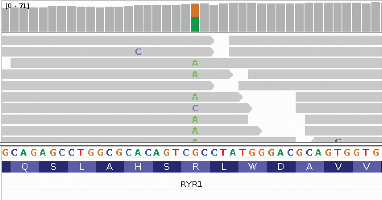
Figure 1.
IGV of the mutation.
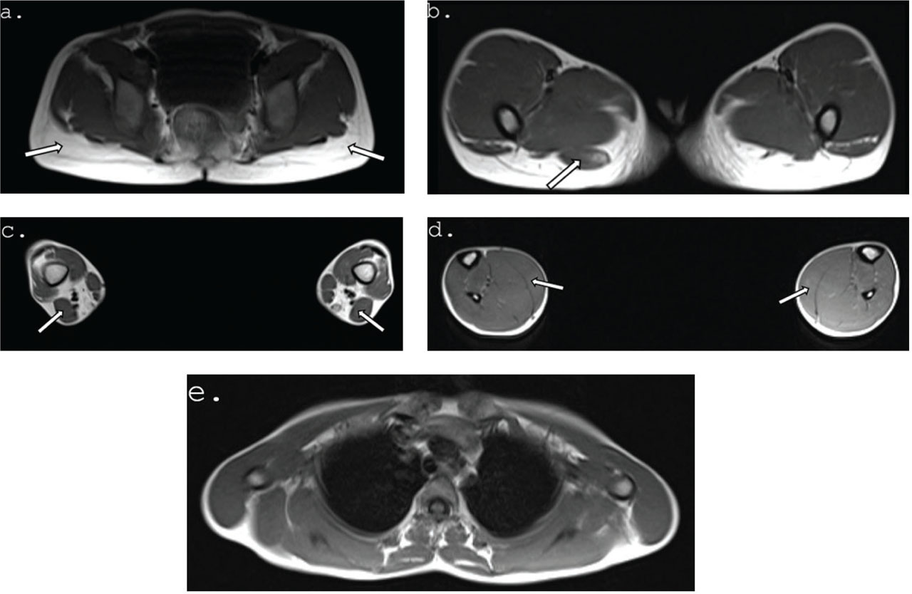
Figure 2.
Magnetic Resonance Imaging (MRI). (A-E) Selected axial T1W images of muscles of patient. (A) Arrows point to complete fatty atrophy in the gluteus maximus muscle bilaterally. (B) Arrows marks relative sparing of the right semitendinosus muscle. (C) Arrows mark hypertrophied short head of biceps femoris muscle bilaterally. (D) Arrows point to relatively hypertrophic soleus and gastrocnemius muscle bilaterally. (E) Normal muscles of the upper girdle.
Table 1.
Clinical features described in published KDS patients and our patients.
| Total number of analyzed cases: | 15 (12 males / 3 females) | (Male) |
|---|---|---|
| Symptom name | Symptom occurrence | Case 1 |
| ptosis | 12 | + |
| lumbar lordosis | 11 | + |
| small build | 10 | + |
| cryptorchidism | 10 | + |
| antimongoloid obliquity | 10 | + |
| low set ears | 10 | + |
| kyphosis/ kyphoscoliosis | 9 | + |
| malignant hyperthermia | 9 | − |
| pectus carinatum/excavatum | 8 | + |
| hypoplastic mandible | 8 | − |
| motor development delay | 8 | + |
| webbed neck | 6 | − |
| high arched palate | 5 | + |
| joint hyperextensibility | 5 | + |
| palpebral slant | 4 | − |
| crowded lower teeth | 2 | − |
| mental development delay | 2 | − |
| scapular winging | 2 | + |
| clinodactyly | 2 | + |
| long face | 2 | − |
| hypertelorism | 2 | + |
| extra fontanelle | 1 | − |
| hip dislocation | 1 | + |
| strabismus | 1 | − |
| pes cavus | 1 | + |
| protuberant nose | 1 | + |
| long upper lip | 1 | + |
| prominent philtrum | 1 | − |
| scaphocephaly | 1 | − |
| rocker bottom foot | 1 | − |
| swallowing problem | 1 | − |
| deep set eyes | 1 | + |