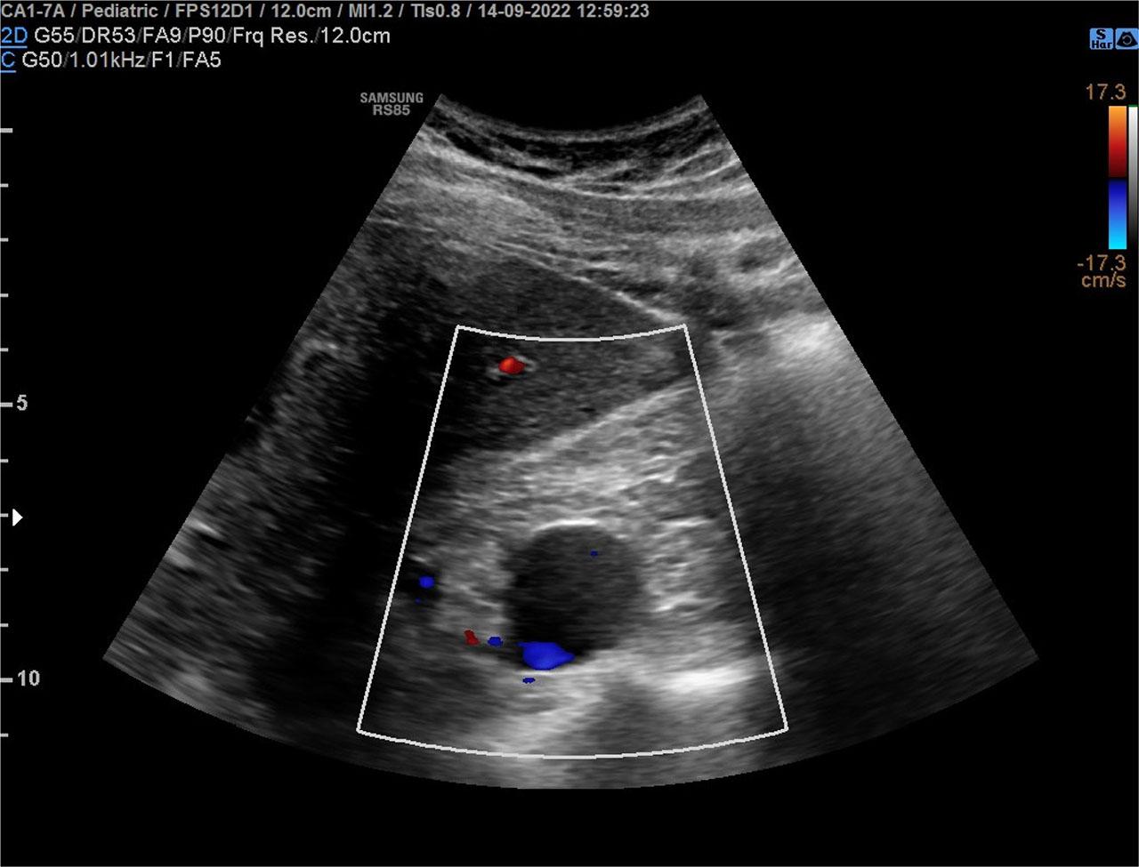
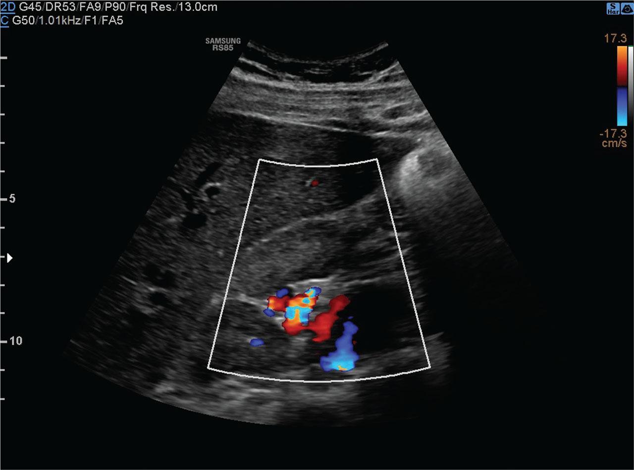
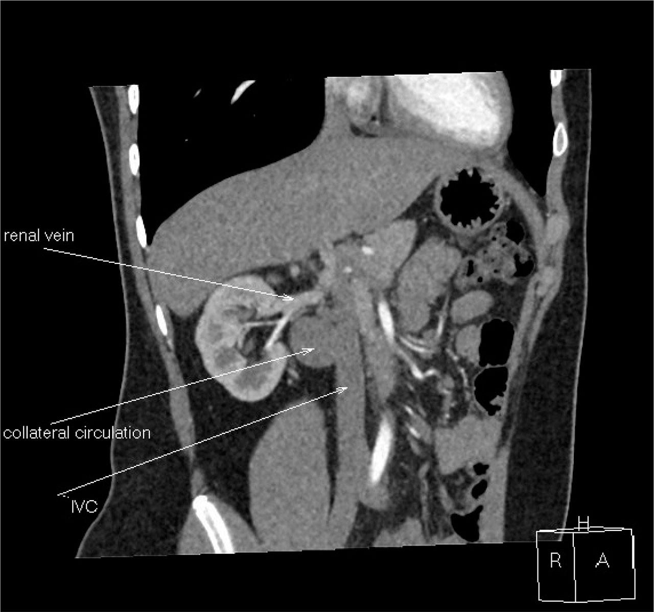
Have a personal or library account? Click to login
IVCA in a Boy with Multilocular Renal Cyst as a Risk Factor for Deep Vein Thrombosis Cover

IVCA in a Boy with Multilocular Renal Cyst as a Risk Factor for Deep Vein Thrombosis

Open Access
|Mar 2025

Figures & Tables

Figure 1.

Abdominal ultrasonography shows multicellular cyst in the hilar region of the right kidney.

Figure 2.

Abdominal ultrasound with power doppler shows blood flow within the right kidney cyst - peripheral circulation.

Figure 3.

CT scan of abdomen: Lack of hepatic and suprarenal IVC, venous outflow leads through vena azygos and hemiazygos.
DOI: https://doi.org/10.34763/jmotherandchild.20242801.d-24-00045 | Journal eISSN: 2719-535X | Journal ISSN: 2719-6488
Language: English
Page range: 146 - 150
Submitted on: Nov 3, 2024
Accepted on: Jan 24, 2025
Published on: Mar 25, 2025
Published by: Institute of Mother and Child
In partnership with: Paradigm Publishing Services
Publication frequency: 1 issue per year

© 2025 Agnieszka Szmigielska, Piotr Skrzypczyk, Michał Szyszka, Magdalena Bukowska, Malwina Wojtas, Aleksandra Jakimów-Kostrzewa, published by Institute of Mother and Child
This work is licensed under the Creative Commons Attribution 4.0 License.