
Table 1
Demographic characteristics of all 18 patients with H-TOF
| Total patients with H-TOF | 18 out anomalies of total oesophageal (4.2%) | |
|---|---|---|
| Age at presentation/referral (n=18) | <1 year | 11 |
| 1–3 years | 4 | |
| 3–6 years | 2 | |
| 6–12 years | 0 | |
| >12 years | 1 | |
| Gender distribution (n=18) | Males | 6 |
| Females | 12 | |
| Number of previous hospitalisations (n=18) | 1 | 8 |
| 2 | 4 | |
| 3 | 5 | |
| >3 | 1 | |
| Associated anomalies (n=6) | ASD | 3 |
| VSD and PDA | 1 | |
| Renal agenesis | 1 | |
| Radial agenesis | 1 |
Table 2
Overlapping reasons for previous hospitalisations in all patients with H-TOF
| Presenting symptoms | No. of patients | |
|---|---|---|
| Respiratory symptoms | Recurrent pneumonias | 18 |
| Bouts of coughing and choking after feeds | 16 | |
| Cyanosis | 8 | |
| Failure to thrive | 14 | |
| Abdominal symptoms | Abdominal distension | 6 |
| Constipation | 8 |
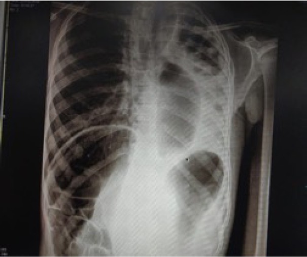
Figure 1
X-ray chest and abdomen of one patient showing complete collapse of the left lung, hyperinflation of the right lung and the Chilaiditi sign.
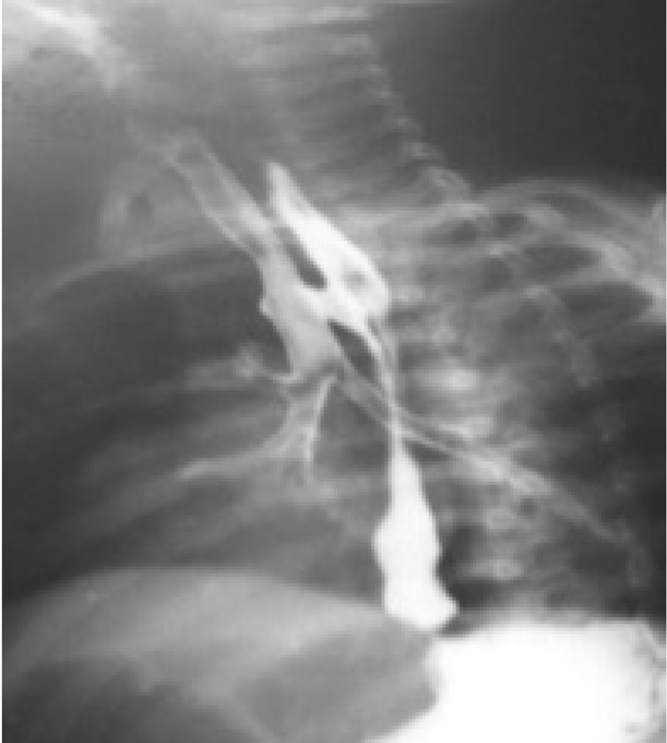
Figure 2
H-TOF identified in the upper gastrointestinal contrast study.
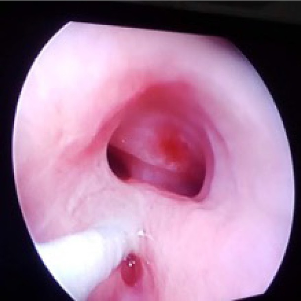
Figure 3
Bronchoscopy showing H-TOF at the C8–T1 level.
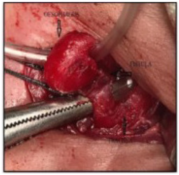
Figure 4
Intraoperative image showing the oesophagus and trachea, as well as identification of the fistula.
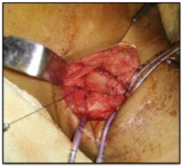
Figure 5
Intraoperative image showing the completed repair.