![Isolation, Identification, and Comprehensive Genomic Characterization of a Bovine Rotavirus G10P[11] Strain in China Cover](https://sciendo-parsed.s3.eu-central-1.amazonaws.com/64727c1c215d2f6c89dc9f62/cover-image.jpg)
Fig. 1.
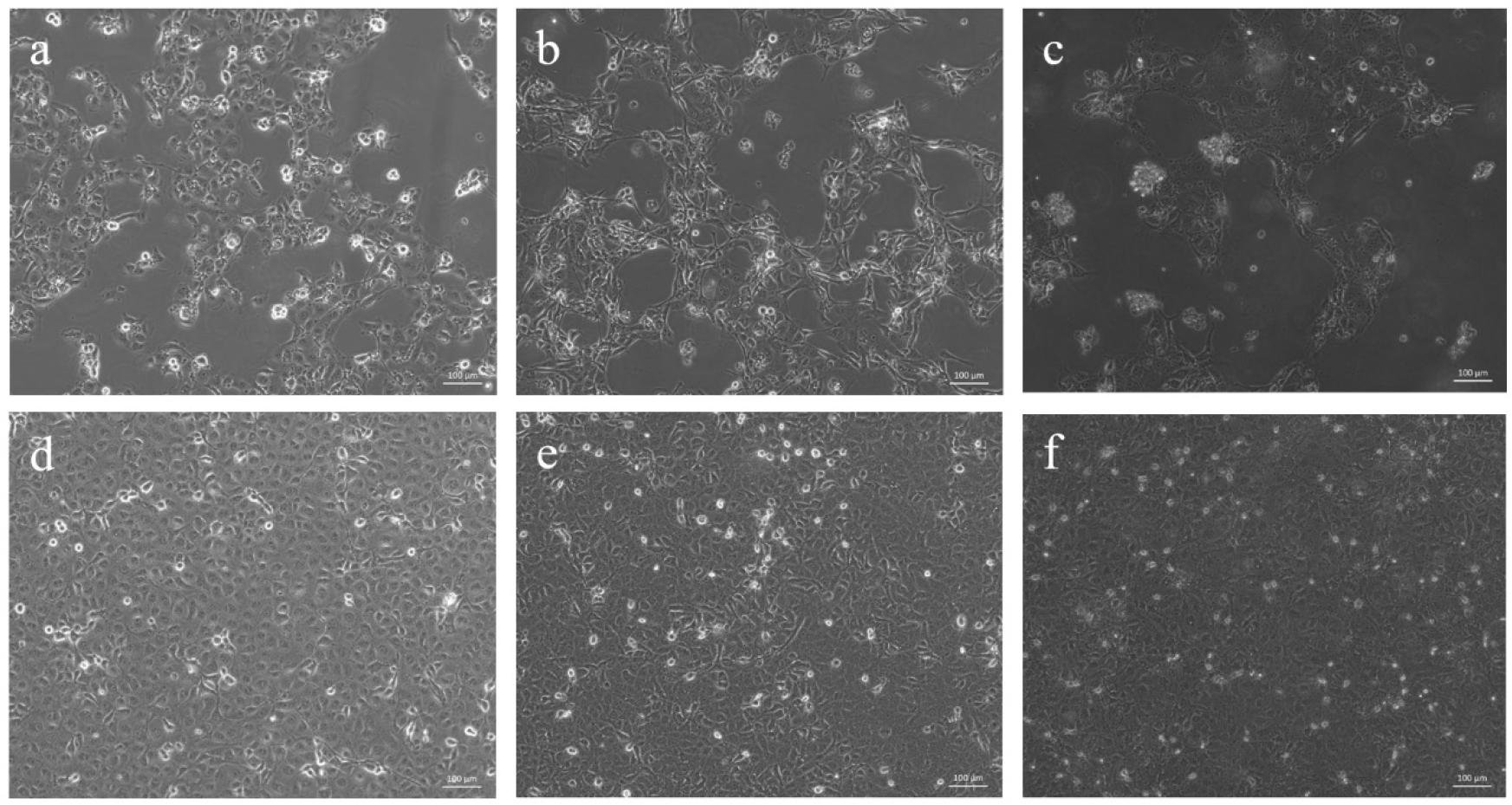
Fig. 2.
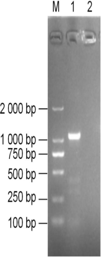
Fig. 3.
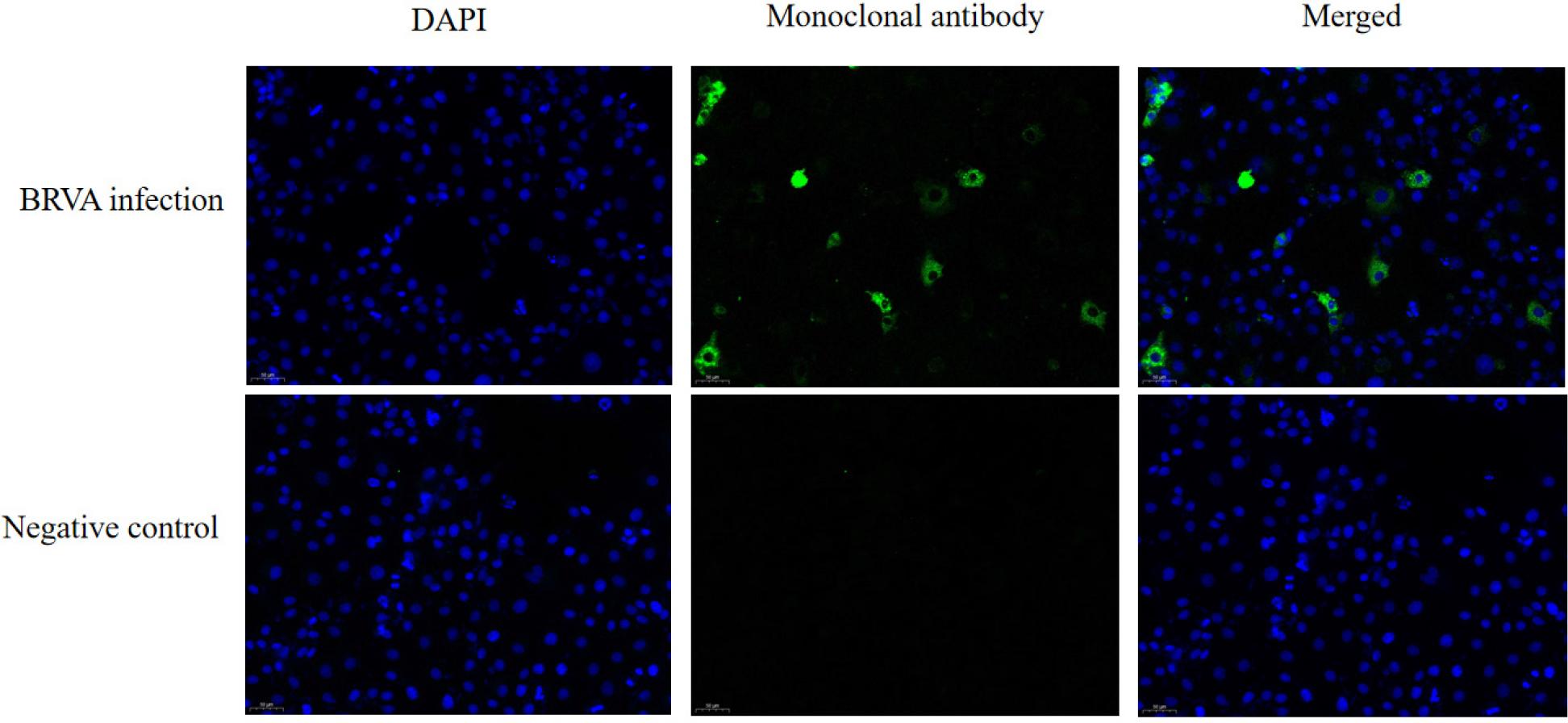
Fig. 4.
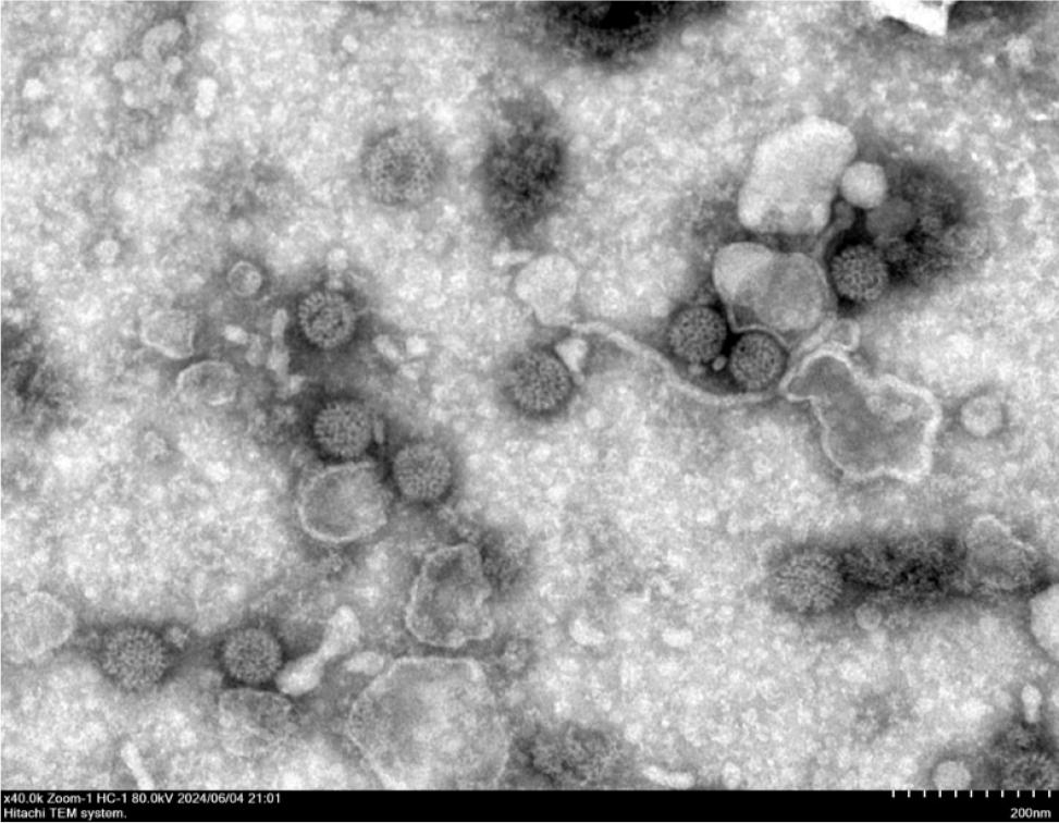
Fig. 5.
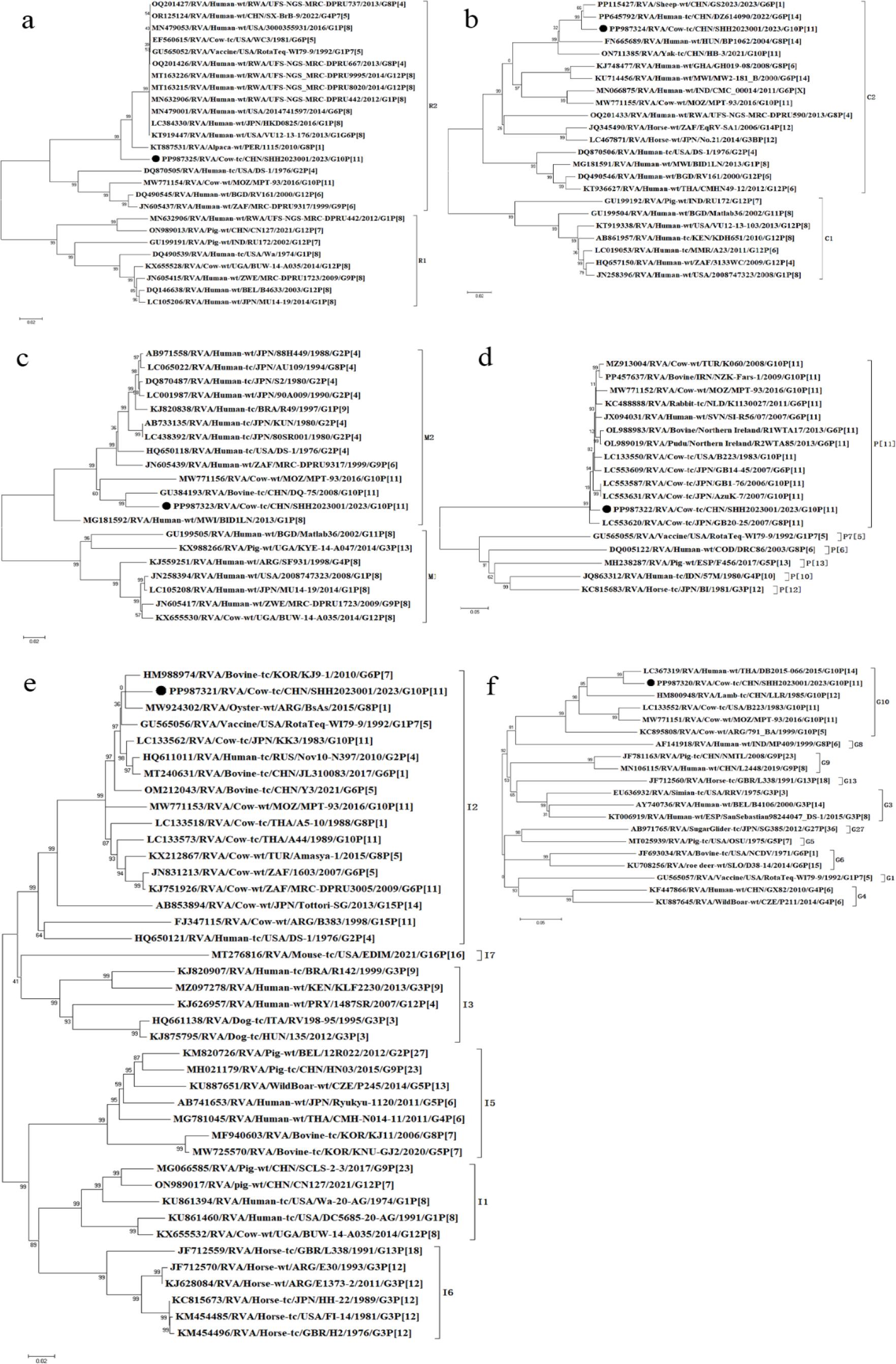
Fig. 6.
Nucleotide sequence identity between strain SHH2023001 and other strains for each gene segment_
| Gene | Closest strain | Accession No. | Homology (%) | Genotype |
|---|---|---|---|---|
| VP7 | RVA/Human-wt/THA/DB2015-66/2015/G10P[14] | LC367319 | 95.41 | G10 |
| VP4 | RVA/Cow-tc/USA/B223/1983/G10P[11] | LC133550 | 96.62 | P[11] |
| VP6 | RVA/Bovine-tc/KOR/KJ9-1/2010/G6P[7] | HM988974 | 97.20 | I2 |
| VP1 | RVA/Human-wt/JPN/HKD0825/2016/G1P[8] | LC384330 | 95.12 | R2 |
| VP2 | RVA/Sheep-wt/CHN/GS2023/2023/G6P[1] | PP115427 | 97.77 | C2 |
| VP3 | RVA/Bovine-tc/CHN/DQ-75/2008/G10P[11] | GU384193 | 94.25 | M2 |
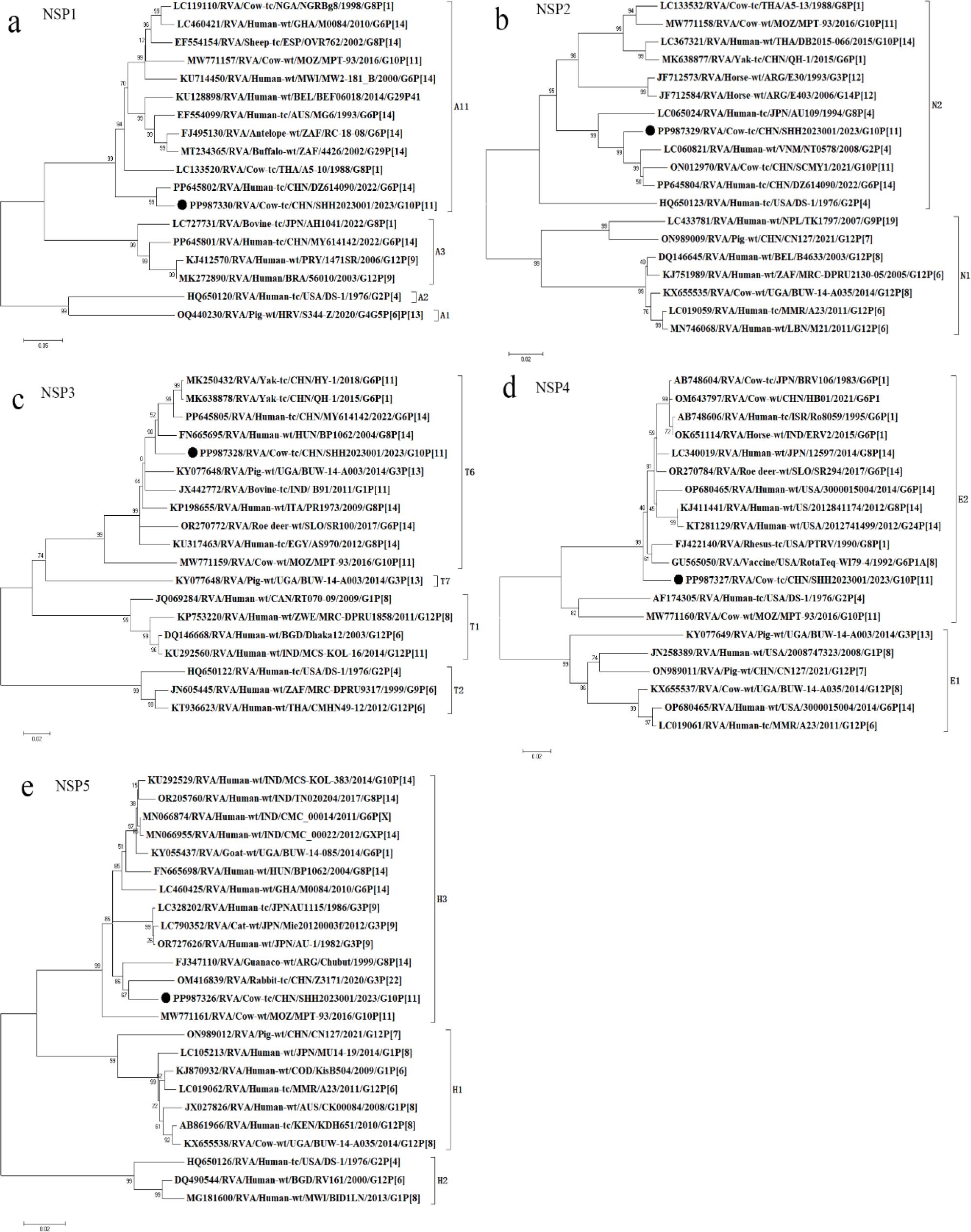
| NSP1 | RVA/Sheep-wt/CHN/GS2023/2023/G6P[1] | PP115432 | 96.47 | A11 |
| NSP2 | RVA/Human-wt/VNM/NT0578/2008/G2P[4] | LC060821 | 96.98 | N2 |
| NSP3 | RVA/Human-wt/HUN/BP1062/2004/G8P[14] | FN665695 | 96.14 | T6 |
| NSP4 | RVA/Horse-wt/IND/ERV2/2015/G6P[1] | OK651114 | 97.44 | E2 |
| NSP5 | RVA/Human-wt/HUN/BP1062/2004/G8P[14] | FN665698 | 97.20 | H3 |