
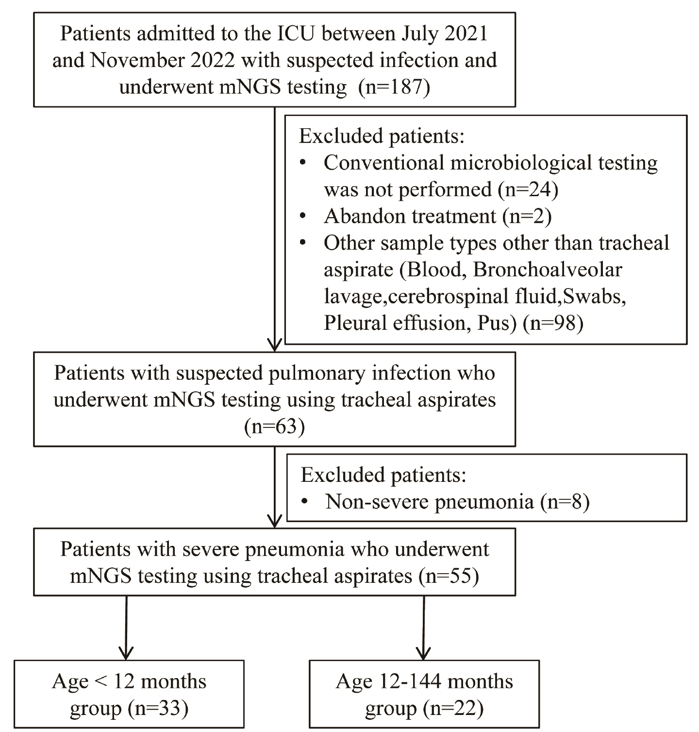
Fig. 1.
Flow diagram of patient inclusion and exclusion.
Table I
Demographic and clinical characteristics of patients.
| Characteristics | Severe pneumonia (n = 55) | Children less than 12 months (n = 33) | Children aged 12~144 months (n = 22) | p-valuea |
|---|---|---|---|---|
| Age, month (M, IQR) | 5.97 (3.28–30.50) | 3.70 (2.80–4.63) | 40.50 (24.75–51.75) | < 0.0001 |
| < 12 | 33 | / | / | / |
| 12–144 | 22 | / | / | / |
| Gender, male, n (%) | 24 (43.64%) | 18 (54.55%) | 6 (27.27%) | 0.0457 |
| Symptoms, n (%) | ||||
| Fever | 24 (43.64%) | 9 (27.27%) | 15 (68.18%) | 0.0027 |
| Cough | 41 (74.55%) | 26 (78.79%) | 15 (68.18%) | 0.3764 |
| Abnormal breathing | 51 (92.73%) | 33 (100%) | 18 (72%) | 0.0012 |
| Unconsciousness | 6 (10.91%) | 3 (9.09%) | 3 (13.64%) | 0.5963 |
| Apathetic | 43 (78.18%) | 23 (69.70%) | 20 (90.91%) | 0.0620 |
| Fidgety | 10 (18.18%) | 2 (6.06%) | 8 (36.36%) | 0.0043 |
| Laboratory examination (mean ± SD) | ||||
| Pulse (times per minute) | 106.9 ± 28.95 | 172.88 ± 22.49 | 143 ± 28.69 | <0.0001 |
| Respiratory rate (times per minute) | 42.31 ± 11.39 | 44.55 ± 9.95 | 38.96 ± 12.77 | 0.0742 |
| WBC | 10.42 ± 4.79 | 11.03 ± 4.52 | 9.49 ± 5.14 | 0.2465 |
| Hb | 105.8 ± 18.72 | 104.82 ± 14.19 | 107.27 ± 24.30 | 0.6382 |
| PLT | 368.13 ± 183.67 | 441.64 ± 187.03 | 257.86 ± 110.54 | 0.0001 |
| NEUT% | 12.38 ± 23.53 | 9.60 ± 17.41 | 16.56 ± 30.53 | 0.2866 |
| LY% | 12.36 ± 23.38 | 17.66 ± 28.21 | 4.42 ± 9.15 | 0.0386 |
| NEUT | 5.02 ± 3.80 | 3.82 ± 2.49 | 6.80 ± 4.71 | 0.0035 |
| HCT | 31.38 ± 7.91 | 31.38 ± 6.79 | 31.38 ± 9.52 | 0.9990 |
| CRP | 21.06 ± 43.04 | 7.35 ± 19.80 | 41.63 ± 58.54 | 0.0029 |
| PCT | 2.61 ± 6.85 | 0.91 ± 2.84 | 5.09 ± 9.78 | 0.0260 |
| BUN | 4.93 ± 4.61 | 4.67 ± 2.95 | 5.33 ± 6.44 | 0.6157 |
| Arterial blood pH | 7.38 ± 0.08 | 7.37 ± 0.08 | 7.40 ± 0.08 | 0.2710 |
| PaO2 | 58.29 ± 17.37 | 57.67 ± 14.03 | 59.23 ± 21.77 | 0.7474 |
| PaCO2 | 43.78 ± 12.70 | 47.33 ± 11.44 | 38.45 ± 12.88 | 0.0097 |
| HCO3− | 26.32 ± 8.26 | 26.93 ± 5.14 | 25.40 ± 11.55 | 0.5058 |
| PaO2/FiO2 | 213.13 ± 64.00 | 221.33 ± 48.89 | 200.82 ± 81.41 | 0.2478 |
| Premature, n | 7 (12.73%) | 6 (18.18%) | 1 (4.55%) | 0.1371 |
| Post operation, n | 3 (5.45%) | 1 (3.03%) | 2 (9.09%) | 0.3322 |
| Intubation, n | 30 (54.55%) | 19 (57.58%) | 11 (50%) | 0.5804 |
| Mechanical ventilation, n | 30 (54.55%) | 19 (57.58%) | 11 (50%) | 0.5804 |
| SOFA score | 3.58 ± 2.03 | 3.48 ± 1.68 | 3.73 ± 2.51 | 0.6691 |
| APACHE II score | 13.45 ± 4.69 | 13.58 ± 3.95 | 13.27 ± 5.72 | 0.8167 |
| LOHS (mean ± SD), day | 16.49 ± 8.83 | 17.39 ± 9.72 | 15.14 ± 7.30 | 0.3579 |
| ICU (mean ± SD), day | 15.24 ± 8.87 | 16.76 ± 9.75 | 12.96 ± 6.97 | 0.1203 |
| Outcomes, n | ||||
| Cured | 53 (96.36%) | 32 (96.97%) | 21 (95.45%) | 0.7687 |
| Died | 2 (3.64%) | 1 (3.03%) | 1 (4.55%) | 0.7687 |
1 ap-Value was the statistically significance result between children aged less than 12 months and 12~144 months. The Student’s t-test was used to compare differences between groups, and the chi-square test was used for analyzing categorical variables. p< 0.05 was considered statistically significant.
1 M – median; IQR – interquartile range; WBC – white blood cell; Hb – hemoglobin; PLT – blood platelet; NEUT – neutrophil cell count; LY – lymphocyte count; HCT – hematocrit; CRP – C-reactive protein; PCT – procalcitonin; BUN – urea nitrogen; PaO2 – partial pressure of oxygen; PaCO2 – partial pressure of carbon dioxide; PaO2/FiO2 – the ratio of PaO2 to fractional inspired oxygen (FiO2); SOFA score – sequential organ failure assessment score; APACHE II score – acute physiology and chronic health evaluation II score; ICU – intensive care unit; LOHS – length of hospital stay.
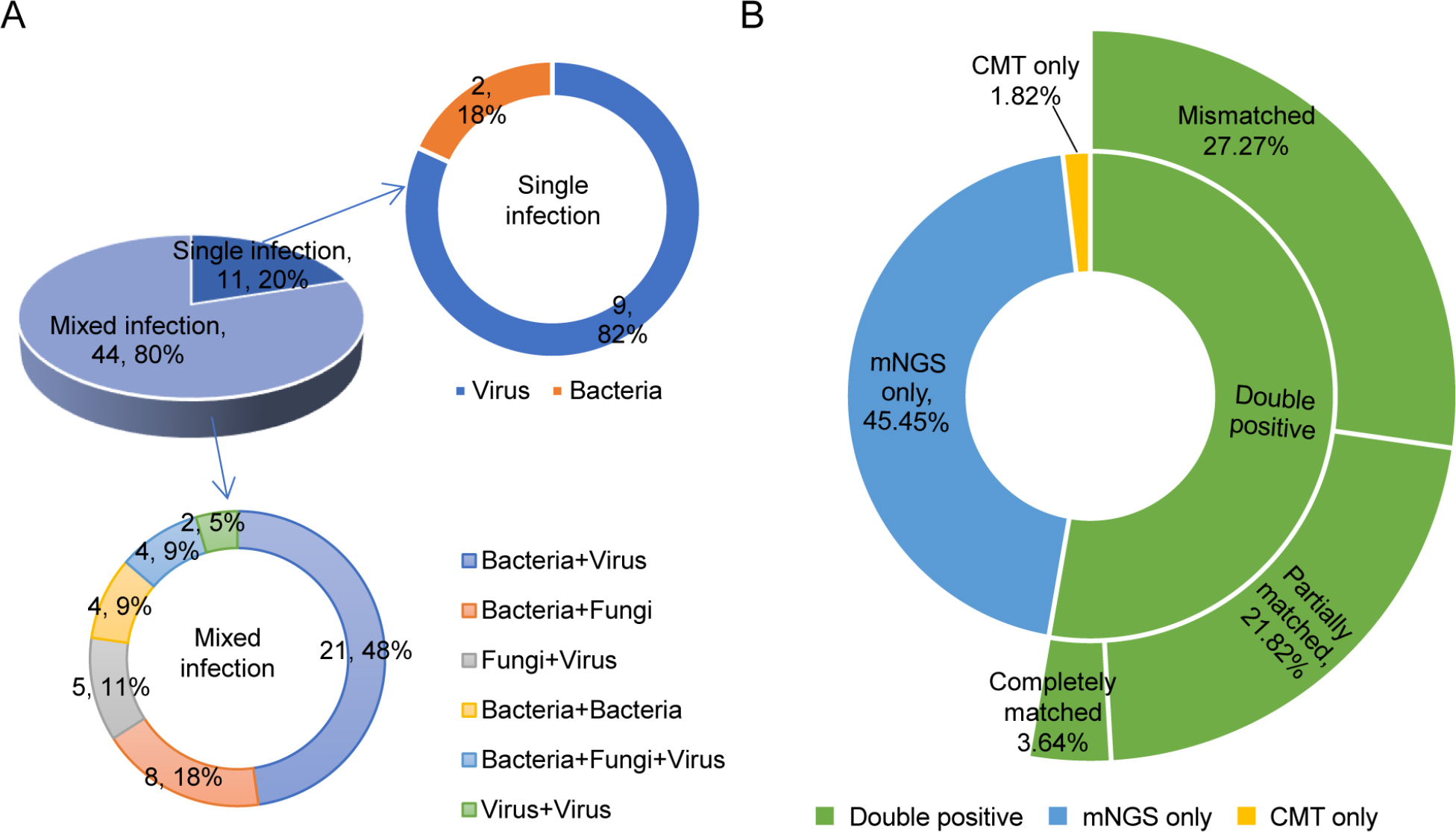
Fig. 2.
The pathogen detected results of mNGS and CMT.
A) The types of infection identified in the children with severe pneumonia. The solid pie chart represents the proportion of single and mixed infections, and the other two hollow pie charts represent the specific pathogen types of single and mixed infections.
B) The coincidence between mNGS and CMT for pathogen identification.
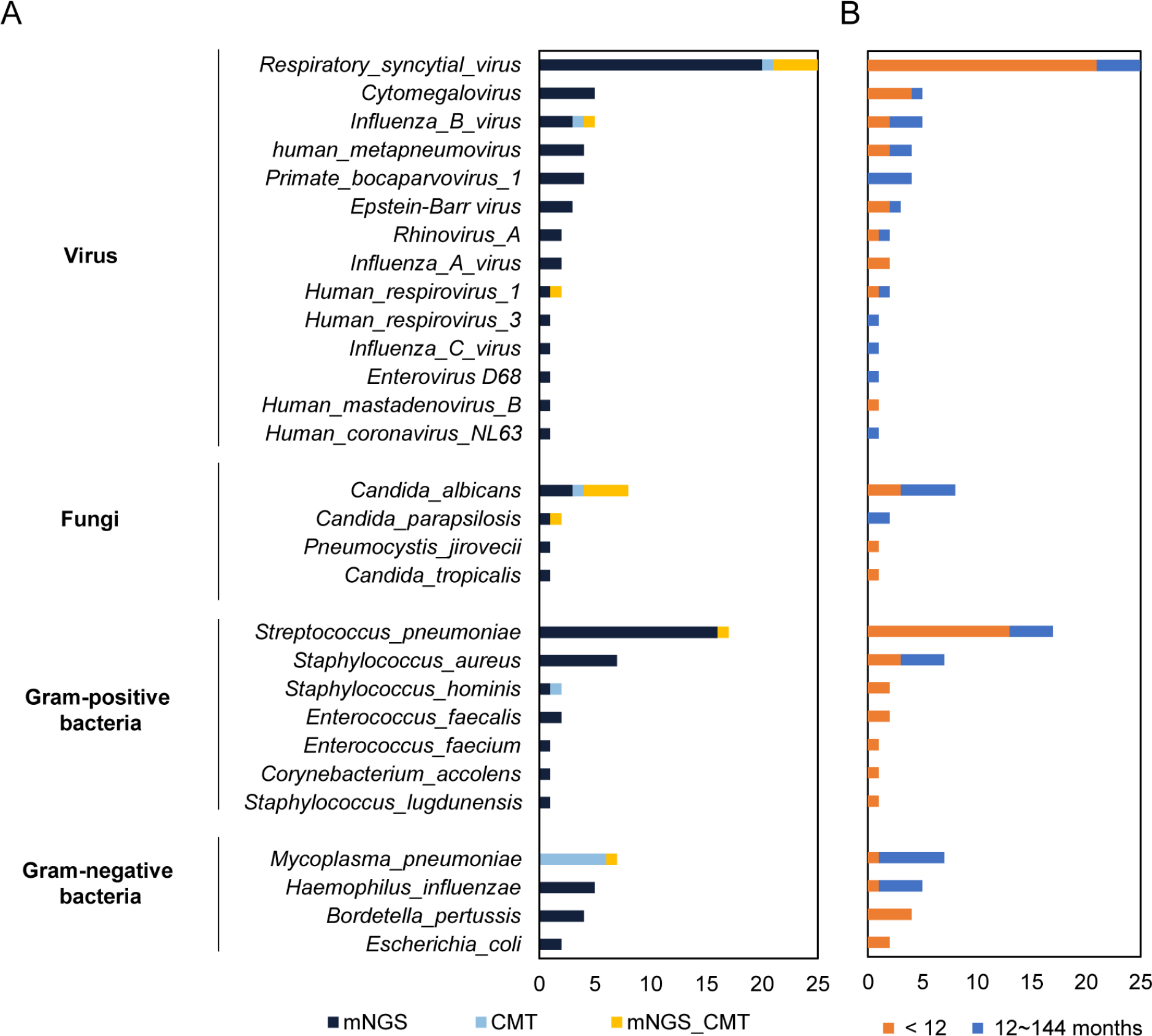
Fig. 3.
The distribution of the identified pathogen spectrum.
A) The distribution of pathogens detected by mNGS and CMT.
B) The distribution of pathogens for children in the < 12 months group and the 12~144 months group.
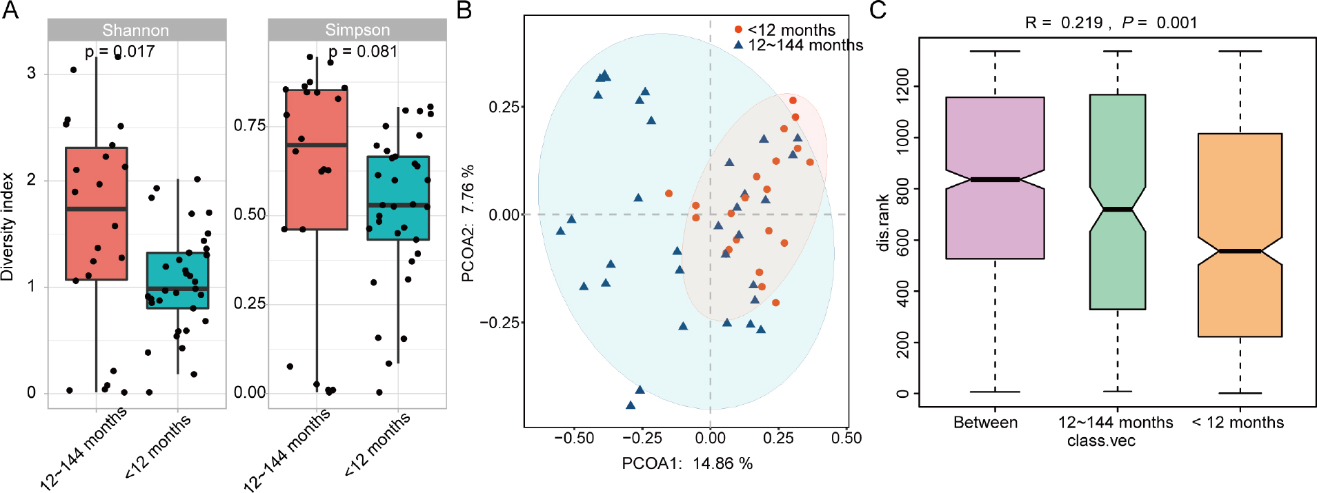
Fig. 4.
Analysis of microbiome alpha and beta diversity between patients < 12 months and 12~144 months.
A) Alpha diversity differences of microbiota communities at the species level between groups. Each point represents one sample from each group. B) PCoA plot is based on Bray–Curtis’s distance between the children in groups. The x-axis is for grouping information, the y-axis is for distance information, the R-value is between (–1, 1) and there are significant differences between groups when the R-value is greater than zero and the P value is less than 0.05.
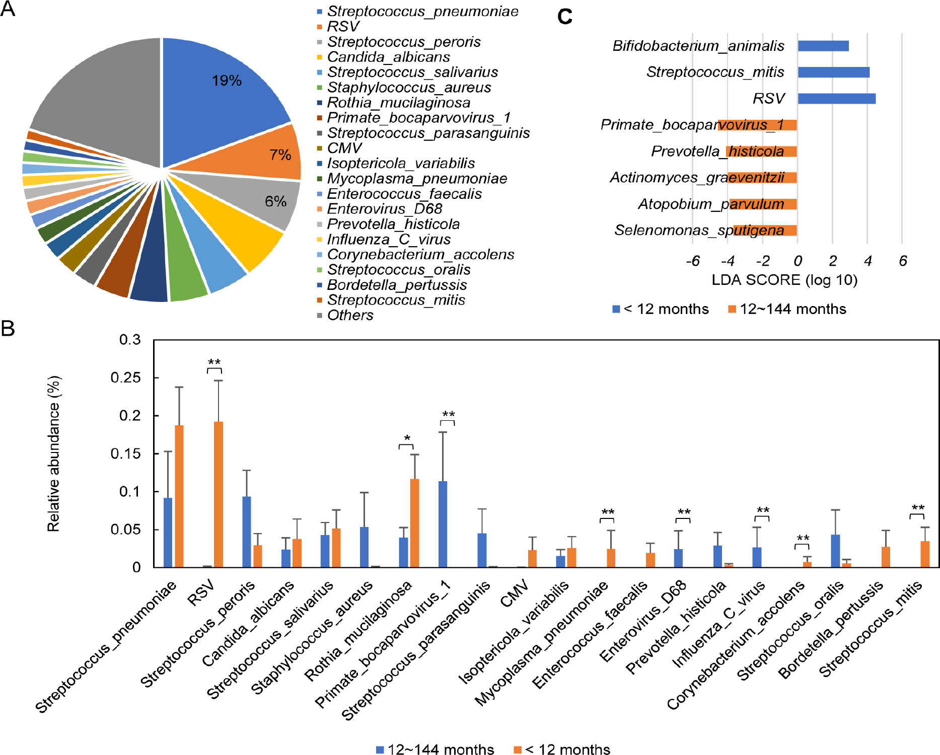
Fig. 5.
The top 20 microorganisms and their correlation with patient clinical data.
A) Top 20 most abundant species and their relative abundance. B) Differences between age groups for the top 20 most abundant microbes. * p-adjust < 0.05; ** p-adjust < 0.01; *** padjust < 0.001. Metastats analysis, p-value was adjusted using the Benjamini and Hochberg method. C) LEfSe analysis of respiratory microbiome between patients < 12 months and 12~144 months. Bar chart showing the log-transformed LDA scores of pathogen species identified by LEfSe analysis (the log-transformed LDA score of 4 as the threshold).
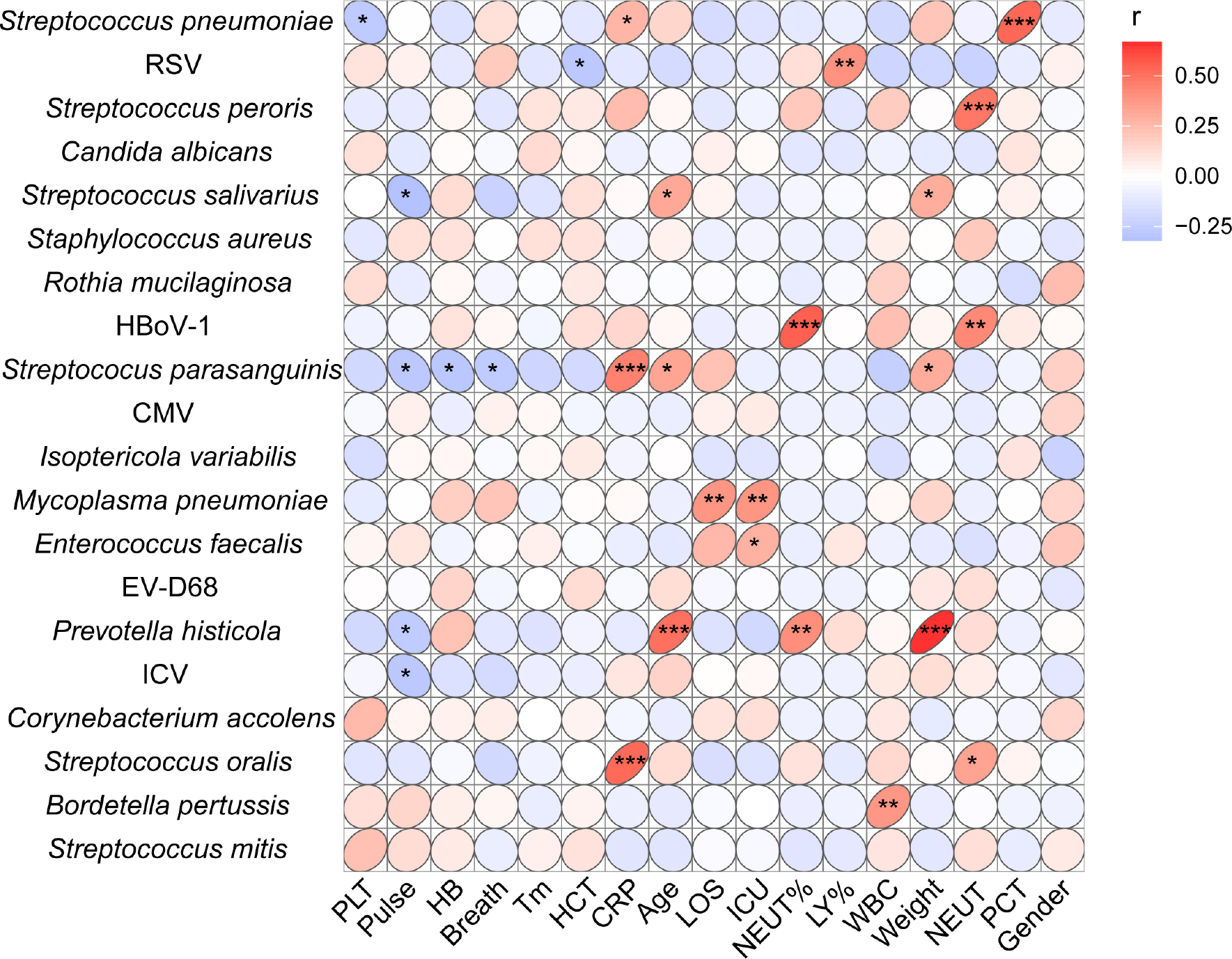
Fig. 6.
Spearman correlations between the top 20 most abundant species and clinical data. * 0.01 < p < 0.05; ** 0.001 < p ≤ 0.01; *** p ≤ 0.001.