
Table I
Primers and crRNA sequences.
| Oligonucleotide | Sequence (5’–3’) |
|---|---|
| RPA forward primers | CCTCTCTTCACGGACCCTCTTTCTACAGC |
| RPA reverse primers | TTCATTCACAAAATTGGGAGCCAGATTGC |
| crRNA | AGUGCCUGGGAGAAAGAUGUCGUUU |
| ssDNA reporters | FAM-TTATTATT-BHQ1 |
| FAM-TTATTATT-Biotin |
Table II
Bacteria and viruses involved in this study.
| Name | Source of strains |
|---|---|
| Shigella flexneri | CMCC(B)51572 |
| Escherichia coli O157:H7 | NCTC12900 |
| Vibrio parahaemolyticus | ATCC® 17802™ |
| Salmonella Typhimurium | Clinical isolates |
| Campylobacter jejuni | ATCC® 33291™ |
| Yersinia enterocolitica | CMCC(B)50024 |
| Human astrovirus | Clinical isolates |
| Rotavirus | ATCC® VR-2274™ |
| Enteric adenovirus | ATCC® VR-930™ |
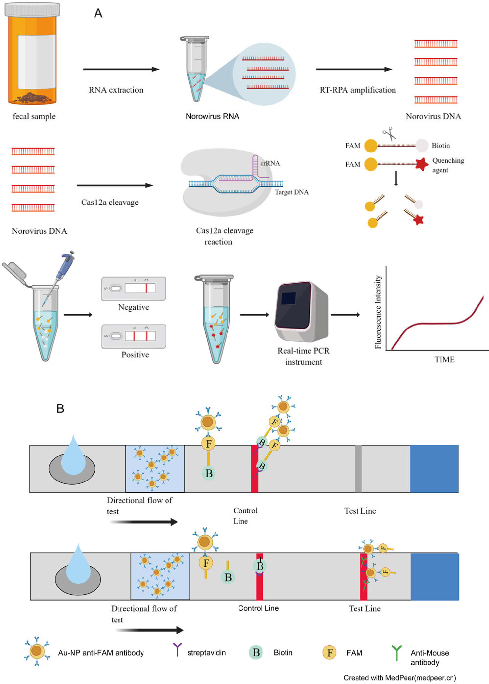
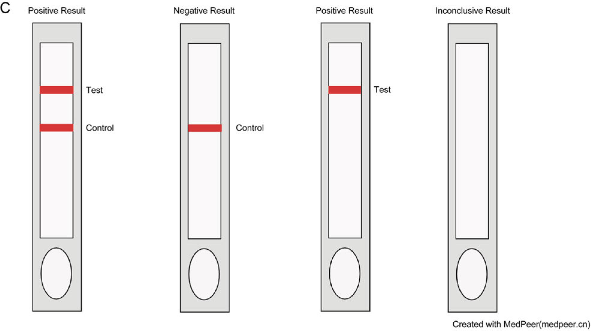
Fig. 1.
Detection principle of RPA-Cas12a-fluorescence analysis and RPA-Cas12a-immunochromatography detection method.
A) RPA-Cas12a method detection process; B) principle of Immunochromatography Flow Strip. C) the schematic diagram for judging the results of immunochromatography flow strips
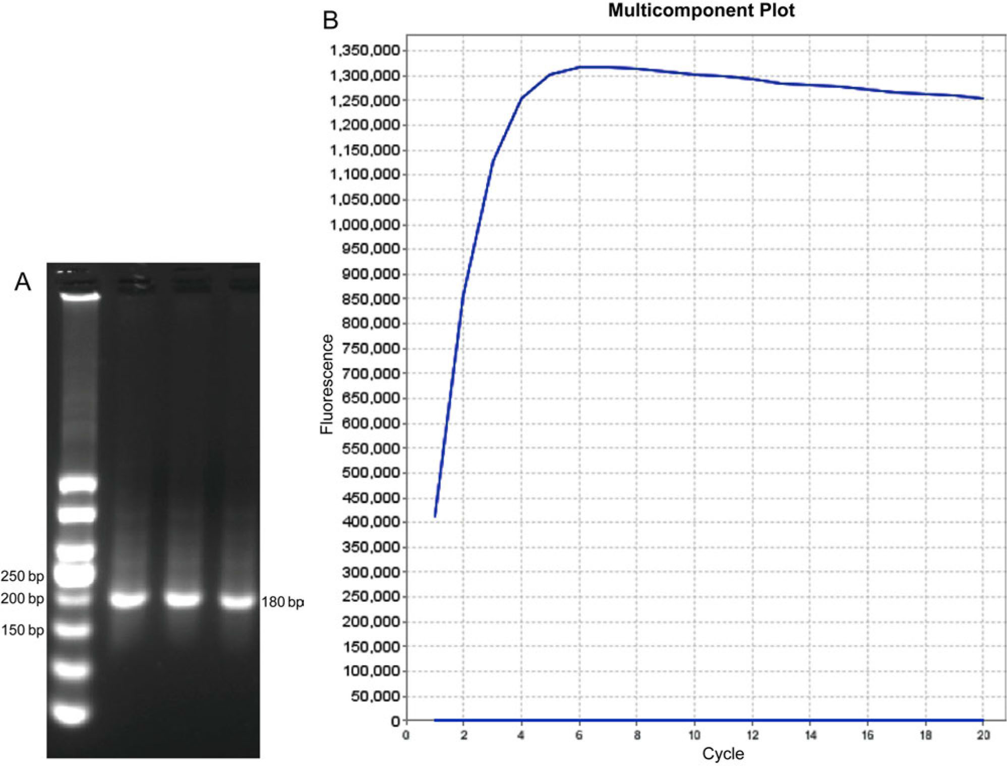
Fig. 2.
RPA amplified fragments and design results of crRNA.
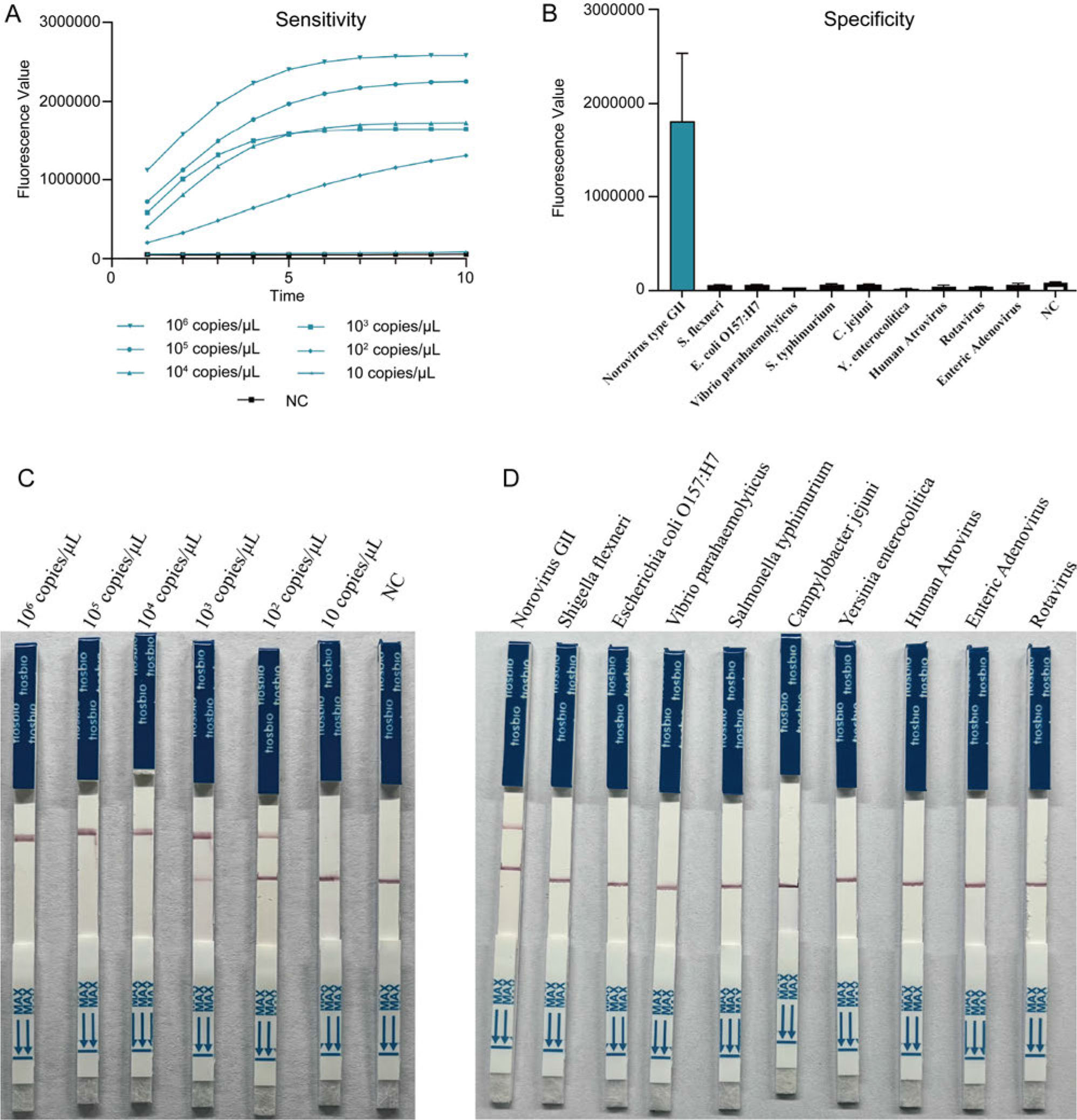
Fig. 3.
Verification of sensitivity and specificity of the RPA-Cas12a-fluorometric and RPA-Cas12a-immunochromatographic assays. A) Sensitivity curves of different concentrations of norovirus positive quality control products amplified by RPA; B) specificity of RPA-Cas12a-fluorometric assay; C) RPA-Cas12a-immunochromatographic results of different concentrations of Norovirus; D) specificity of RPA-Cas12a-immunochromatographic assay
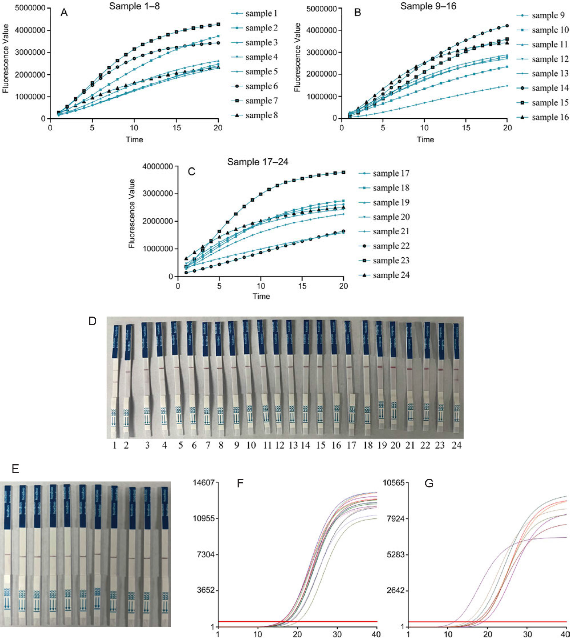
Fig. 4.
Detection of GII norovirus in clinical samples based on CRISPR-Cas12a.
A, B, C) Positive results of RPA-Cas12a-fluorescence assay of 24 samples; D) positive results of RPA-Cas12a-immunochromatographic assay of 24 samples; E) negative results of RPA-Cas12a-immunochromatographic assay of 11 samples; F, G) qPCR results of 24 positive samples