
Table I
Background information on the participants.
| Number of participants | 10 |
| Age (median/minimum-maximum) | 8/2–12 |
| Male/female | 7/3 |
| Atopic dermatitis | 8 |
| Bronchial asthma | 7 |
| Allergic rhinitis | 8 |
| IgE (total) IU/ml (25%–75%) | 1,157 (384–2,744) |
| IgE (milk) kUA/l (25%–75%) | 50 (27–66) |
| EASI score (25%–75%) | 0.3 (0.05–2.1) |
| Serum TARC (pg/ml) (25%–75%) | 551 (333–976) |
[i] IgE – Immunoglobulin E,
[ii] EASI – Eczema Area and Severity Index,
[iii] TARC – Thymus and Activation-Regulated Chemokine
Table II
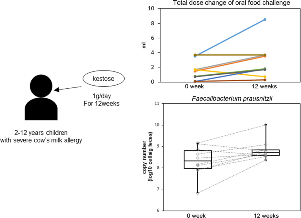
Oral food challenge results for all patients.
| First OFC | Second OFC | |||||
|---|---|---|---|---|---|---|
| TD (ml) | TS (points) | TS/Pro | TD (ml) | TS (points) | TS/Pro (points/g) | |
| Patient 1 | 3.5 | 10 | 94 | 8.5 | 5 | 19 |
| Patient 2 | 1.5 | 25 | 550 | 3.5 | 25 | 236 |
| Patient 3 | 1.7 | 15 | 291 | 3.7 | 1 | 9 |
| Patient 4 | 1.7 | 31 | 601 | 0.7 | 20 | 943 |
| Patient 5 | 0.1 | 10 | 3300 | 1.8 | 0 | 0 |
| Patient 6 | 0.8 | 16 | 660 | 1.8 | 20 | 367 |
| Patient 7 | 3.7 | 15 | 134 | 3.7 | 15 | 134 |
| Patient 8 | 0.1 | 10 | 3300 | 0.3 | 25 | 2750 |
| Patient 9 | 0.7 | 11 | 519 | 1.7 | 20 | 388 |
| Patient 10 | 3.7 | 15 | 134 | 3.7 | 40 | 357 |
[i] OFC – Oral Food Challenge, TD – Total Dose, TS – Total Score of ASCA, Pro – cumulative protein dose
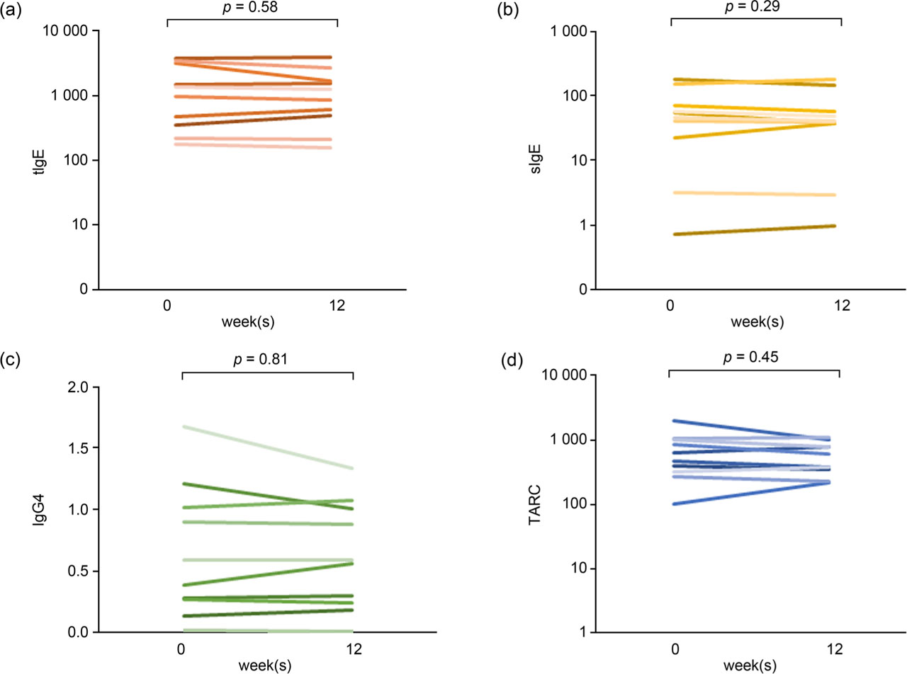
Fig. 1.
Changes in the patients’ immunological parameters.
The laboratory data from 10 participants before and after the study are shown. a) Total Immunoglobulin E (tIgE), b) milk-specific Immunoglobulin E (sIgE), c) casein-specific Immunoglobulin G4 (IgG4), d) Thymus and Activation-Regulated Chemokine (TARC). No significant changes were seen after 12 weeks (p > 0.05, Wilcoxon's signed-rank test).
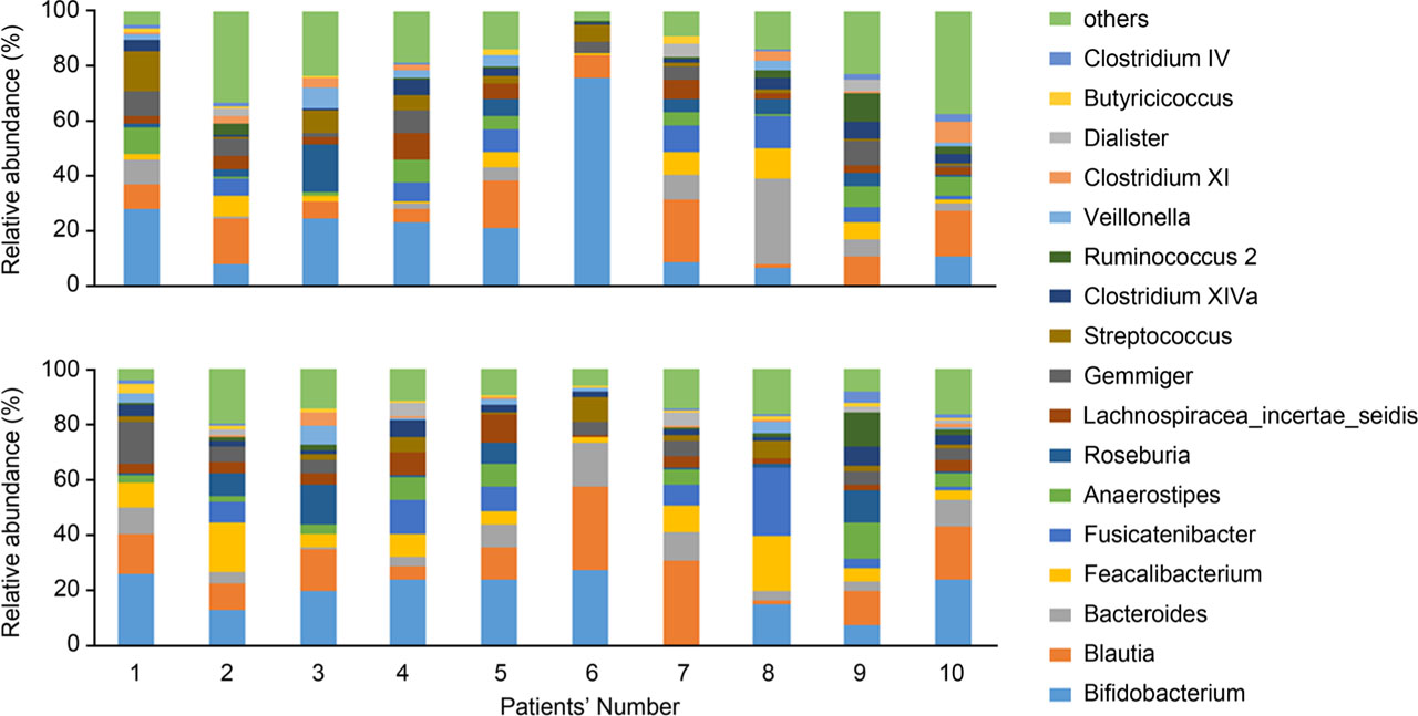
Fig. 2.
Relative abundance of gut microbiota at 0 and 12 weeks.
This bar graph shows the composition of the intestinal microbiota in each of the 10 subjects. The numbers on the horizontal axis of the graphs represent the patient number, and the graphs in the same column represent the results for the same subject. The results for the top 17 genera with the highest occupancy are shown. The top row shows the results at the beginning of the study, and the bottom row shows the results 12 weeks into the study.
Table III
Relative abundance of gut microbiota.
| 0w | 12w | p-value* | |
|---|---|---|---|
| Median (25%–75%) | Median (25%–75%) | ||
| xBifidobacterium | 16.2 (8.3–24.2) | 21.7 (13.3–24.1) | 0.72 |
| Blautia | 9.5 (6.8–16.9) | 13.6 (10.0–22.7) | 0.24 |
| Bacteroides | 4.0 (0.8–8.3) | 6.6 (3.7–9.8) | 0.20 |
| Faecalibacterium | 3.8 (1.4–7.0) | 6.8 (4.4–9.7) | 0.01 |
| Fusicatenibacter | 6.0 (0.3–8.1) | 5.3 (0.4–8.8) | 0.29 |
| Anaerostipes | 5.0 (1.0–7.7) | 4.4 (2.3–7.3) | 0.39 |
| Roseburia | 3.8 (0.5–5.3) | 0.8 (0.4–8.0) | 0.88 |
| Lachnospiracea_incertae_sedis | 3.0 (2.7–5.3) | 3.8 (2.5–4.5) | 0.65 |
| Gemmiger | 4.6 (1.0–7.5) | 4.7 (1.1–5.6) | 0.67 |
| Streptococcus | 2.3 (0.9–6.5) | 2.4 (1.6–4.7) | 0.88 |
| Clostridium XlVa | 2.9 (1.1–4.5) | 2.2 (1.8–3.9) | 0.24 |
| Ruminococcus2 | 0.6 (0.4–3.1) | 0.8 (0.3–1.7) | 0.51 |
| Veillonella | 1.8 (0.2–3.4) | 0.8 (0.2–3.2) | 0.96 |
| Clostridium XI | 1.4 (0.1–3.0) | 0.6 (0.4–0.9) | 0.33 |
| Dialister | 0.2 (0.0–2.2) | 0.9 (0.0–1.9) | 0.59 |
| Butyricicoccus | 0.4 (0.1–1.3) | 0.9 (0.6–1.6) | 0.20 |
| Clostridium IV | 0.6 (0.1–1.5) | 0.3 (0.2–1.0) | 0.45 |
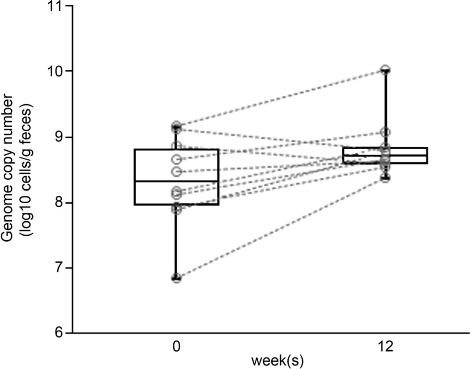
Fig. 3.
Absolute quantification of intestinal microbiota (qPCR).
The logarithm of the genome copy number of Faecalibacterium prausnitzii per gram of feces is shown at baseline (0w) and after 12 weeks of intervention (12w). Dotted lines connect changes in the same participants. Eight of 10 patients exhibited nominal increases from 0 to 12 weeks, but this difference was not statistically significant (p > 0.05, Wilcoxon's signed-rank test).