
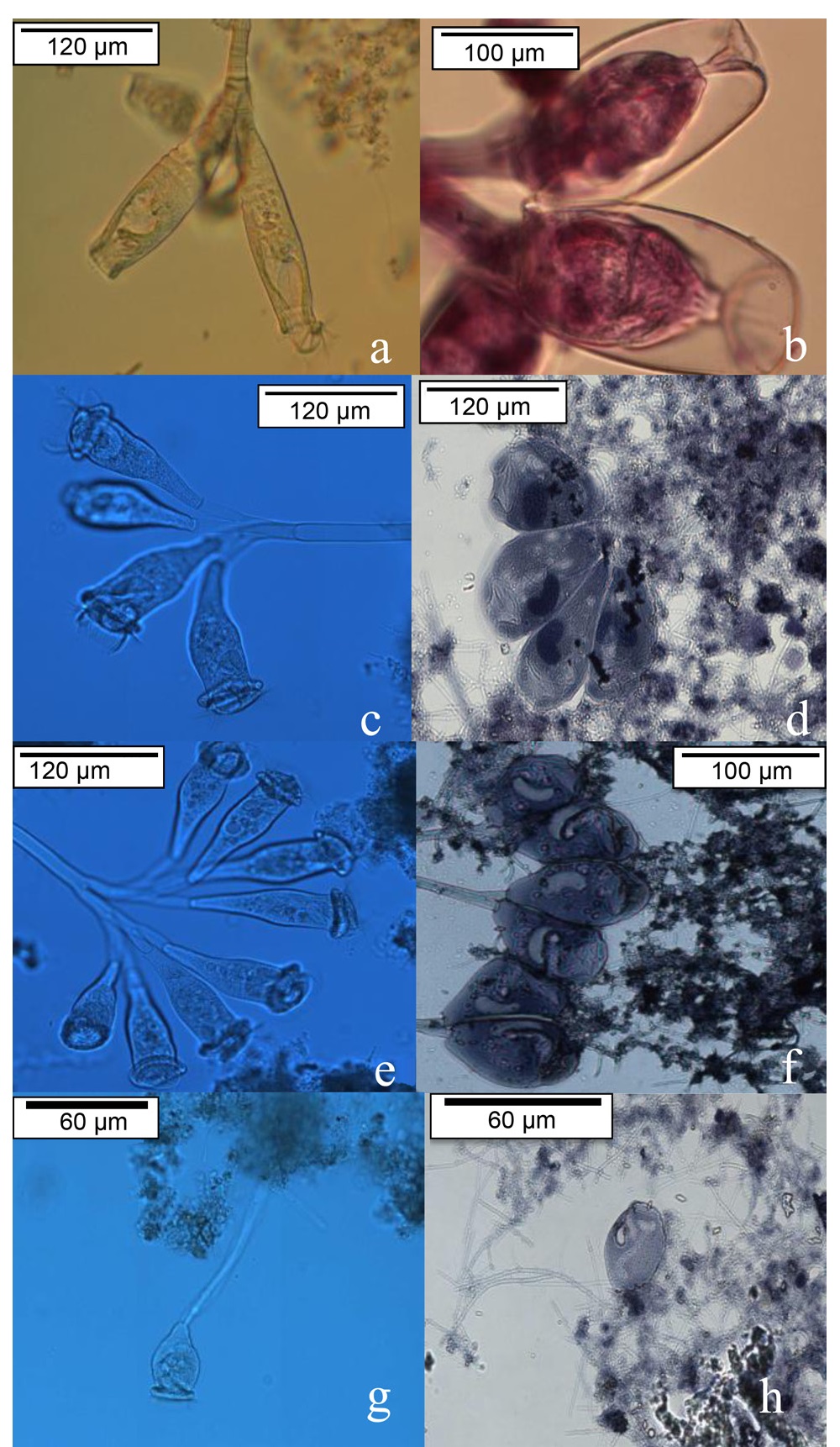
Fig. 1
Micrographs of colonial peritrichous protozoa.
a) Fresh sample and b) stained with Harris hematoxylin technique at 40x; c) 4-zoid fresh colony using Nomarski technique and d) FD staining at 40x; e) 8-zoid fresh colony using Nomarski technique and f) the MFD staining at 40x; and g) fresh samples of Vorticella campanula and h) stained with the MFD technique (“J” shaped nucleus) at 20x magnification.
Table I
The density of peritrichous protozoa (peritrichous protozoa × 103/ml).
| Time (days) | Zoothamnium paraentzii | Epistylis plicatilis | Vorticella campanula | Epistylis rotans | Carchesium polypinum | Vorticella convallaria | Vorticella aquadulcis | Total (× 103density protozoa/ml) |
|---|---|---|---|---|---|---|---|---|
| 1 | 11 | 8 | 10 | 7 | 33 | 1.5 | 1 | 72 |
| 10 | 142 | 34 | 57 | 47 | 17 | 2 | 2 | 299 |
| 20 | 267 | 34 | 57 | 47 | 17 | 2 | 2 | 425 |
| 30 | 220 | 92 | 96 | 76 | 11 | 5 | 2 | 500 |
| 40 | 244 | 63 | 76 | 61 | 14 | 3 | 2 | 462 |
| 60 | 232 | 77 | 86 | 68 | 12 | 4 | 2 | 481 |
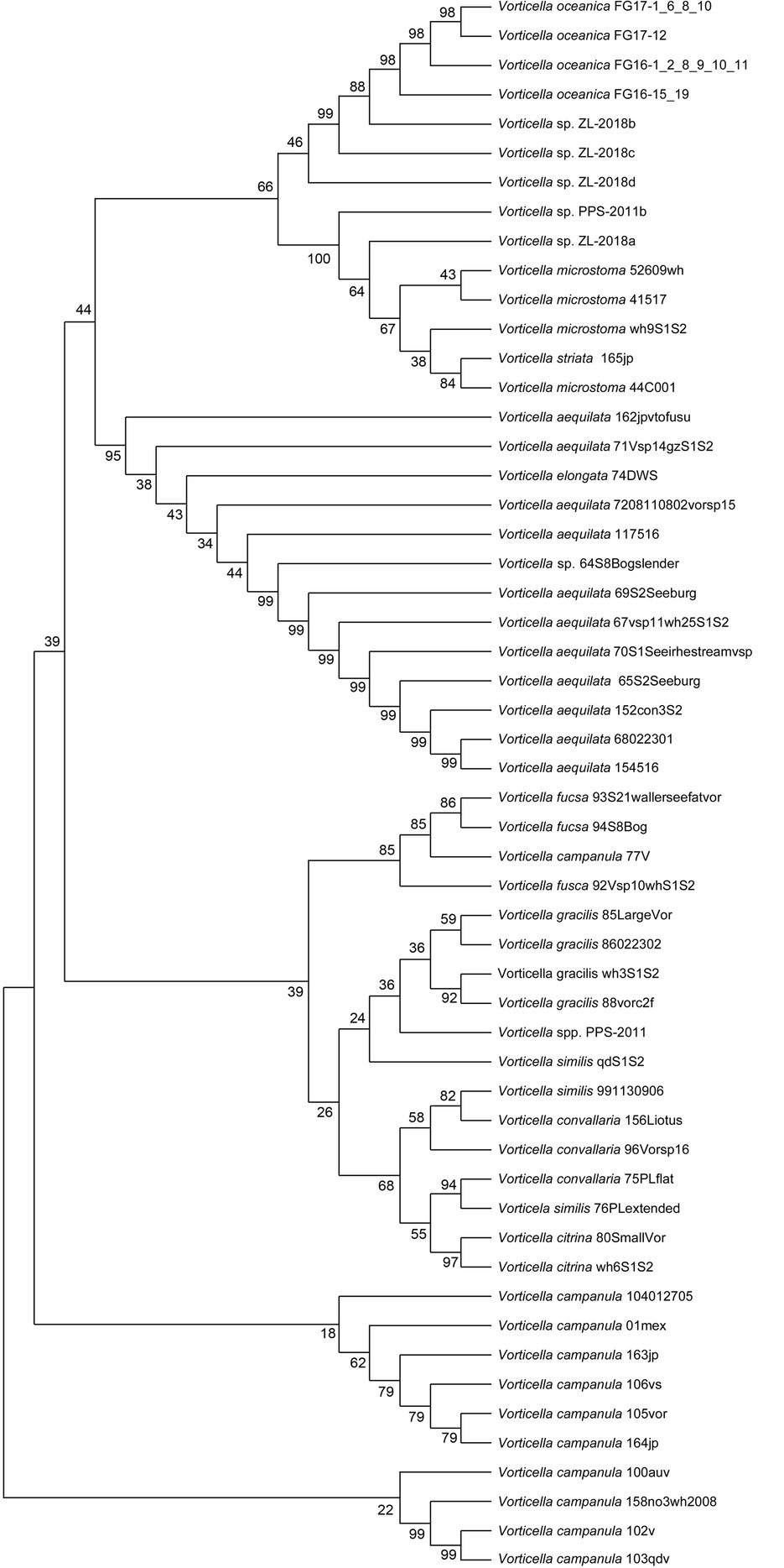
Fig. 2
Identification of Vorticella campanula 01mex by a phylogenetic analysis based on its ITS 18S rRNA sequence.
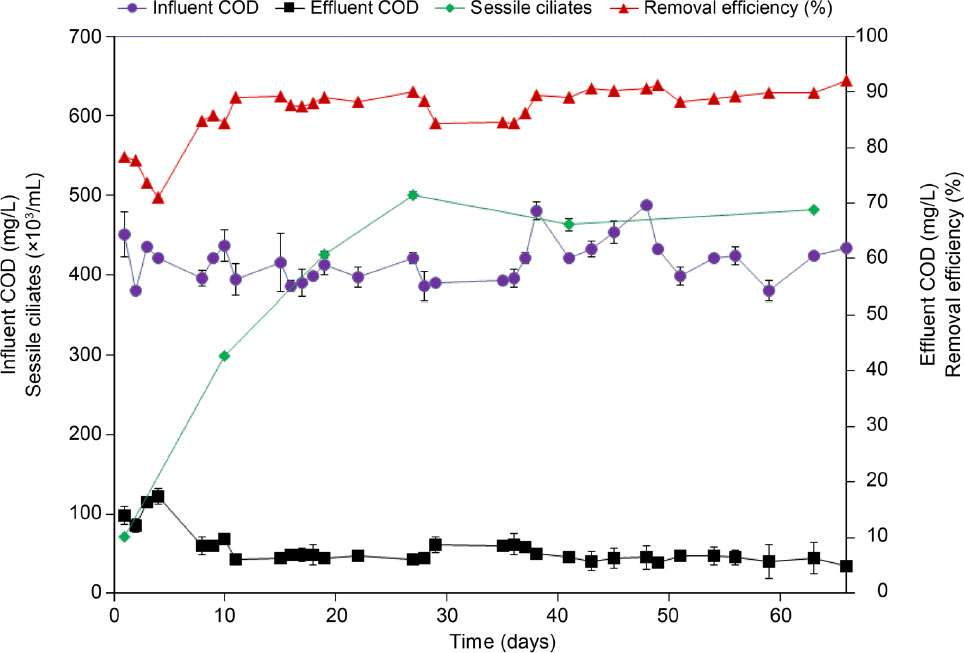
Fig. 3.
Relationship between protozoan density and COD in the SBR.  Protozoa density;
  Influent COD;
  Effluent COD;
  Removal efficiency (%).
Table II
The mean abundance of peritrichous protozoan species.
| Species | Average density (protozoa × 103/ml) |
|---|---|
| Zoothamnium paraentzii | 185.79a |
| Epistylis plicatilis | 51.18b |
| Vorticella campanula | 63.56b |
| Epistylis rotans | 50.79b |
| Carchesium polypinum | 17.31c |
| Vorticella convallaria | 2.85d |
| Vorticella aquadulcis | 1.64d |
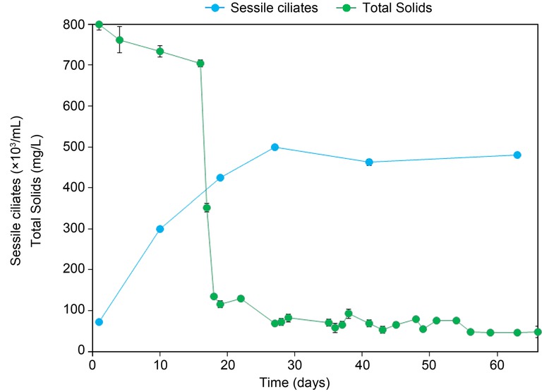
Fig. 4
Behavior of sessile ciliates with respect to the concentration of total solids.
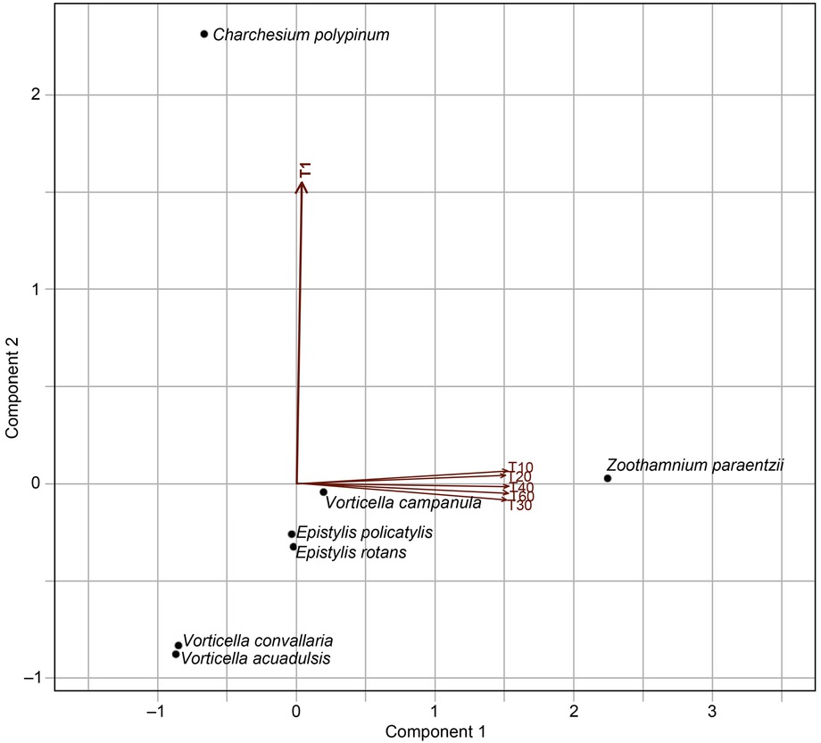
Fig. 5
Principal Component Analysis (PCA): relationship between the abundance of protozoa and other microorganisms, time, and removal percentage. Axes 1 and 2 account for 54.9% and 22.1% of the total variation presented, respectively.
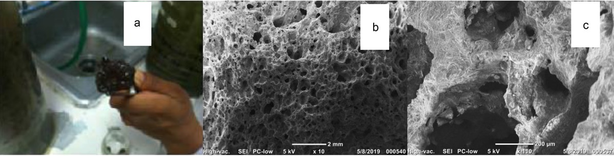
Fig. 6
Properties of tezontle as supporting material of the SBR. a) A piece of tezontle containing adhered peritrichous colonies; b) Scanning Electron Microscopy (SEM) of tezontle stones; c) closeup of tezontle pores.