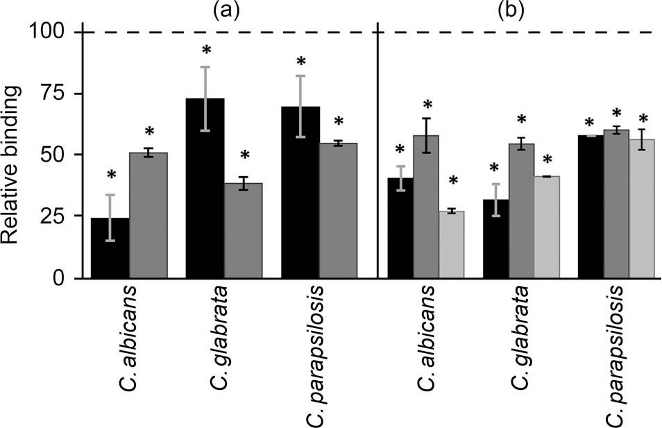
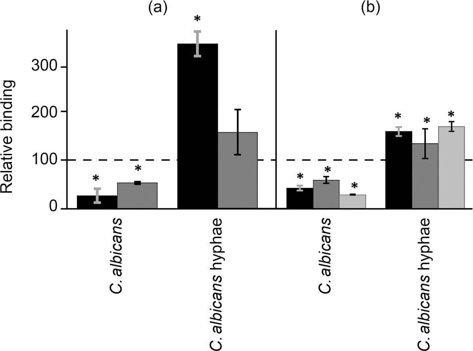
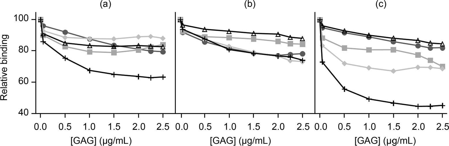
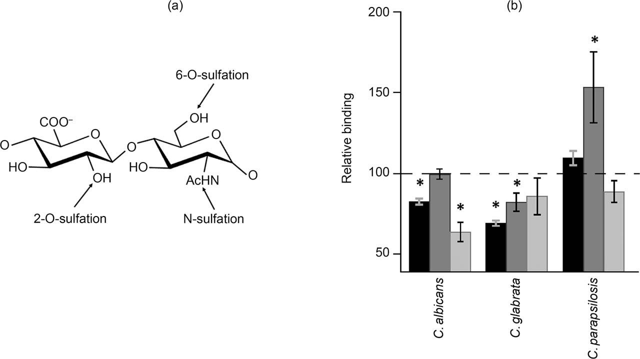
Have a personal or library account? Click to login
Cell Surface Glycosaminoglycans as Receptors for Adhesion of Candida spp. to Corneal Cells Cover

Cell Surface Glycosaminoglycans as Receptors for Adhesion of Candida spp. to Corneal Cells

Open Access
|Mar 2022

Figures & Tables

Fig. 1

Effect of reduction of cell GAGs on fungal adhesion to corneal epithelial cells. a) Inhibition of fungal adhesion to HCE-2 cells treated with rhodamine B (black bars) and genistein (gray bars). b) Inhibition of fungal adhesion to HCE-2 cells treated with heparinases I and III (black bars), chondroitinase ABC (dark gray bars), or a mix of heparinases I and III + chondroitinase ABC (gray bars). Data were normalized using the adhesion values of fungi to non-treated cells, which were given the arbitrary value of 100. Error bars represent standard deviations. Statistically significant differences are denoted by *, which indicates p < 0.05.

Fig. 2

Effect of reducing cell GAGs on the adhesion of C. albicans yeast cells and hyphal cells to corneal epithelial cells. a) Inhibition of fungal adhesion to HCE-2 cells treated with rhodamine B (black bars) and genistein (gray bars). b) Inhibition of fungal adhesion to HCE-2 cells treated with heparinases I and III (black bars), chondroitinase ABC (dark gray bars), or a mix of heparinases I and III + chondroitinase ABC (gray bars). Data were normalized using the adhesion values of fungi adhesion to non-treated cells, given the arbitrary value of 100. Error bars represent standard deviations. Statistically significant differences are denoted by *, which indicates p < 0.05.

Fig. 3

Inhibition of fungal adhesion to corneal epithelial cells by the presence of different GAGs. a) adhesion of C. albicans, b) C. glabrata, c) C. parapsilosis in the presence of different concentrations of HS (

), CSA (

), CSB (

), CSC (

) and a mixture of all GAGs (+). Data were normalized using the adhesion values of fungi to non-treated cells, which was given the arbitrary value of 100.
Inhibition of fungal adhesion to corneal epithelial cells by the presence of different GAGs. a) adhesion of C. albicans, b) C. glabrata, c) C. parapsilosis in the presence of different concentrations of HS ( ), CSA ( ), CSB ( ), CSC ( ) and a mixture of all GAGs (+). Data were normalized using the adhesion values of fungi to non-treated cells, which was given the arbitrary value of 100.

Fig. 4

Influence of specific N- and O-sulfations on pathogen adherence to corneal epithelial cells. a) Structure of an HS disaccharide unit showing the specific positions of sulfations. b) Inhibition of fungal adhesion to corneal epithelial cells with silencing of the genes involved in N-sulfation (dark bars), 2-O-sulfation (dark gray bars), and 6-O-sulfation (gray bars). Data were normalized using the adhesion values of fungi to non-treated cells, which was given the arbitrary value of 100. Error bars represent standard deviations. Statistically significant differences are denoted by *, which indicates p < 0.05.
DOI: https://doi.org/10.33073/pjm-2022-008 | Journal eISSN: 2544-4646 | Journal ISSN: 1733-1331
Language: English
Page range: 55 - 62
Submitted on: Dec 13, 2021
Accepted on: Feb 10, 2022
Published on: Mar 14, 2022
Published by: Polish Society of Microbiologists
In partnership with: Paradigm Publishing Services
Publication frequency: 4 times per year

© 2022 Helena Ordiales, Ignacio Alcalde, Fernando Vázquez, Jesús Merayo-Lloves, Luis M. Quirós, Carla Martín Cueto, published by Polish Society of Microbiologists
This work is licensed under the Creative Commons Attribution-NonCommercial-NoDerivatives 4.0 License.