
Table I
PCR primers used in this study.
| Genes (reference) | Primer names and sequences (5′-3′) | Size of nested PCR products (bp) |
|---|---|---|
| ITS (Lee et al. 1998) | 1724F 5′-AAGTTGATCAAATTTGGTC-3′ ITS2R 5′-CTCGGACGAGGATCCTCGCC-3′ ITS1F 5′-CGTAGGTGAACCTGCGGAAGGATC-3′ ITS2R1 5′-GTTCAGCGGGTGATCCTGCCTG-3′ | 578 |
| sod (Esteves et al. 2010b) | MnSOD_Fw 5′-GGGTTTAATTAGTCTTTTTAGGCAC-3′ MnSOD_Rw 5′-CATGTTCCCACGCATCCTAT-3′ SODF3 5′-AGTCTTTTTAGGCACTTGAACCT-3′ SODR4 5′-TCCAAGAATAACTTTGCCTTGAGT-3′ | 560 |
| dhfr (Lane et al. 1997) | FR208 5′-GCAGAAAGTAGGTACATTATTACGAGA-3′ FR1018 5′-AAGCTTGCTTCAAACCTTGTGTAACGCG-3′ FR242 5′-GTTTGGAATAGATTATGTTCATGGTGTACG-3′ FR1038 5′-GCTTCAAACCTTGTGTAACGCG-3′ | 798 |
| dhps (Ma et al. 1999) | DHPS1 5′-CAAATTAGCGTATCGAATGACC-3′ DHPS2 5′-GCAAAATTACAATCAACCAAAGTA-3′ DHPS3 5′-AGCGCCTACACATATTATGG-3′ DHPS4 5′-GTTCTGCAACCTCAGAACG-3′ | 278 |
| cyb (Esteves et al. 2010a) | CytbFw 5′-CCCAGAATTCTCGTTTGGTCTATT-3′ CytbRw 5′-AAGAGGTCTAAAAGCAGAACCTCAA-3′ CytbF3 5′-TCTCGTTTGGTCTATTGGTG-3′ CytbR4 5′-AAGCAGAACCTCAAATTCAAGATA-3′ | 590 |
| mtLSU rRNA (Wakefield 1996) | pAZ102_E 5′-GATGGCTGTTTCCAAGCCCA-3′ pAZ102_H 5′-GTGTACGTTGCAAAGTACTC-3′ pAZ102_X 5′-GTGAAATACAAATCGGACTAGG-3′ pAZ102_Y 5′-TCACTTAATATTAATTGGGGACC-3′ | 252 |
| β-Tub (Pasic et al. 2020) | Pneumo Tub_F 5′-TCATTAGGTGGTGGAACGGG-3′ Pneumo Tub_R 5′-ATCACCATATCCTGGATCCG-3′ | 303 |
| 26S rRNA (Pasic et al. 2020) | PneumoLSU_F 5′-TCAGGTCGAACTGGTGTACG-3′ PneumoLSU_R 5′-TGTTCCAAGCCCACTTCTT-3′ | 297 |
Table II
Nucleotide polymorphic sites and number of plasmid clones sequenced at eight distinct loci of Pneumocystis jirovecii.
| Locus | Genotypesa | Locationb | No. of plasmid clones sequenced | ||
|---|---|---|---|---|---|
| SX_0001 | SX_0002 | SX_0003 | |||
| ITS | ITS 4 | KC470776 | 0 | 12 | 0 |
| ITS 10 | JQ365725 | 0 | 0 | 4 | |
| ITS 16 | AB469817 | 0 | 0 | 8 | |
| ITS 22 | KC470795 | 6 | 0 | 0 | |
| ITS 59 | MK300661 | 10 | 0 | 0 | |
| sod | sod 1 | 110C/215T | 11 | 13 | 8 |
| sod 2 | 110T/215C | 0 | 0 | 2 | |
| dhps | dhps WT | 165A (55Thr) / 171C (57Pro) | 12 | 12 | 12 |
| dhps | dhfr312 | 312C (117Gly) | 12 | 11 | 11 |
| cyb | cyb 1 | 279C/348A/516C/547C/566C/838C | 0 | 0 | 6 |
| cyb 2 | 279C/348A/516C/547C/566C/838T | 0 | 8 | 0 | |
| cyb 7 | 279C/348A/516C/547C/566T/838C | 9 | 0 | 0 | |
| cyb 8 | 279T/348A/516C/547C/566C/838C | 0 | 0 | 3 | |
| mt LSU rRNA | mt1 | 85C/248C | 0 | 0 | 2 |
| mt2 | 85A/248C | 0 | 0 | 8 | |
| mt3 | 85T/248C | 10 | 10 | 0 | |
| β-Tub | β-Tub 1 | 86G/281A | 8 | 0 | 6 |
| β-Tub 2 | 86G/281G | 4 | 12 | 5 | |
| 26S rRNA | 26S 2 | 86T/290A | 12 | 11 | 0 |
| 26S 3 | 86C/290A | 0 | 0 | 5 | |
| 26S 4 | 86A/290A | 0 | 0 | 6 | |
ITS – internal transcribed spacer regions of rRNA operon, sod – superoxide dismutase, dhfr – dihydrofolate reductase, dhp – dihydropteroate synthase, WT – wild-type, cyb – cytochrome b, mt – mitochondrial large rRNA subunit, β-Tub – β-tubulin, 26S rRNA – 26S ribosomal RNA gene
b – the genotype locations according to the studies previously reported (Walker et al. 1998; Ma et al. 1999; Beard et al. 2000; Denis et al. 2000; Takahashi et al. 2002; Esteves et al. 2010b; Maitte et al. 2013; Xue et al. 2019; Pasic et al. 2020)
Table III
Clinical characteristics of patients with Pneumocystis jirovecii pneumonia.
| Clinical information | Patient No. | ||
|---|---|---|---|
| SX_0001 | SX_0002 | SX_0003 | |
| Age (years) | 65 | 51 | 65 |
| Sex | Male | Male | Male |
| Underlying conditions | NAa | Hepatic cysts | ILD |
| Thoracic HRCT findings | GGO d + | GGO + | GGO + |
| HIV 1/2 antibody | +/– | +/– | –/– |
| CD4 T-lymphocyte count (cells/µl) | 232 | 176 | NA |
| Serum parameters | |||
| 1,3-β-D-glucan, normal < 10 pg/ml | > 600 | NA | > 600 |
| Lactate dehydrogenase, normal 120–250 U/l | 432 | 699 | 9,734 |
| C-reactive protein, normal 0–6 mg/l | 73.63 | 129.17 | 340.00 |
| Procalcitonin, normal 0–0.05 ng/ml | 0.975 | 0.161 | 11.26 |
| Partial pressure of oxygen, normal 80–110 mmHg | 80 | 65 | 59.70 |
| Erythrocyte sedimentation rate, normal 0–15 mm/h | 61.10 | 60.80 | 47.30 |
| Concurrent infection | – | – | C.n. and B.c.b |
| Anti-PCP therapya | – | – | – |
| HAART before PCP | – | – | – |
| Clinical outcomes | survived | survived | died |
NA – not available; ILD – interstitial lung disease; HRCT – high-resolution computed tomography; GGO – ground-glass opacity; HIV – human immunodeficiency virus; HAART – highly active antiretroviral therapy
+ – positive, − – negative
Table IV
Genotypes of Pneumocystis jirovecii detected at eight genetic loci.
| Patient No. | HIV1/2 anti-body | Genotypes at 8 loci | |||||||
|---|---|---|---|---|---|---|---|---|---|
| ITS | sod | dhfr | dhps | cyb | mtLSU rRNA | β-Tub | 26S rRNA | ||
| SX_0001 | +/− | ITS2 + ITS59 | sod1 | dhfr312 | WT | cyb7 | mt3 | β-Tub1 + β-Tub2 | 26S2 |
| SX_0002 | +/− | ITS4 | sod1 | dhfr312 | WT | cyb2 | mt3 | β-Tub2 | 26S2 |
| SX_0003 | −/− | ITS10 + ITS16 | sod1 + sod2 | dhfr312 | WT | cyb1 + cyb8 | mt1 + mt 2 | β-Tub1 + β-Tub2 | 26S3 + 26S4 |
[i] ITS – internal transcribed spacer regions of rRNA operon, sod – superoxide dismutase, dhfr – dihydrofolate reductase, dhps – dihydropteroate synthase, WT – wild-type, cyb – cytochrome b, mt – mitochondrial large rRNA subunit, β-Tub – β-tubulin, 26S rRNA – 26S rRNA gene
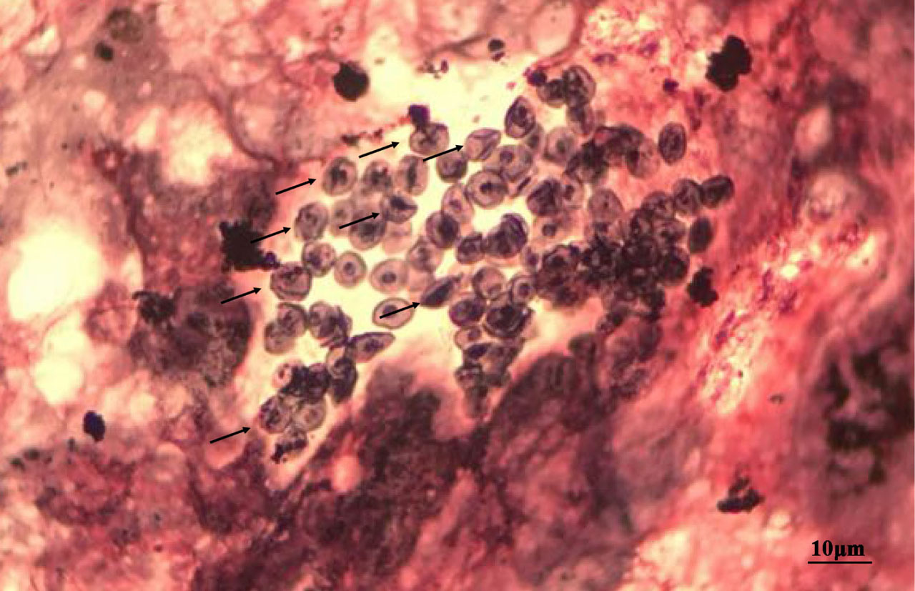
Fig. 1
Identification of Pneumocystis jirovecii using GMS staining methods. Cysts appear as brown or puce spheres or ovoids with a small black stick inside (arrows). The reddish background instead of the typical greenish background is most likely due to periodic acid treatment and without light green counterstaining in our staining method.
Table V
Multi-locus sequence type (MLST) profiles of P. jirovecii from PCP patients in this study in comparison with known P. jirovecii MLST profiles in Fungal MLST Database.
| MLST types* | β-Tub | cyb | 26S rRNA | sod | Patient no. |
|---|---|---|---|---|---|
| 3 | 1 | 1 | 4 | 2 | SX_0003 |
| 8 | 2 | 8 | 4 | 1 | SX_0003 |
| 13 | 1 | 1 | 4 | 1 | SX_0003 |
| 15 | 1 | 8 | 4 | 1 | SX_0003 |
| 19 | 1 | 8 | 3 | 2 | SX_0003 |
| 21 | 2 | 1 | 3 | 1 | SX_0003 |
| 22 | 2 | 1 | 3 | 2 | SX_0003 |
| 23 | 2 | 1 | 4 | 1 | SX_0003 |
| 35 | 2 | 7 | 2 | 1 | SX_0001 |
| 51 | 1 | 7 | 2 | 1 | SX_0001 |
| 52 | 2 | 2 | 2 | 1 | SX_0002 |
| NA | 1 | 1 | 3 | 2 | SX_0003 |
| NA | 2 | 8 | 3 | 1 | SX_0003 |
| NA | 2 | 8 | 3 | 2 | SX_0003 |
| NA | 2 | 1 | 4 | 2 | SX_0003 |
| NA | 2 | 8 | 4 | 2 | SX_0003 |
| NA | 1 | 1 | 3 | 1 | SX_0003 |
| NA | 1 | 8 | 3 | 1 | SX_0003 |
| NA | 1 | 8 | 4 | 2 | SX_0003 |
* – The first 11 MLST types (numbered 3 to 52) identified in this study correspond to those in the Fungal MLST Database at http://mlst.mycologylab.org
NA – types identified in this study and not available from the Fungal MLST Database
In both patients SX_0001 and SX_0003 (with co-infection of two genotypes at 2 or 6 loci, respectively), there were a total of four and 64 potential MLST profiles, respectively. Only two and 16 of those profiles are listed in this table while the true profiles could not be determined in this study.