Introduction
Microbes play a prominent and important role in the plant-microbe ecosystem (Hodge and Fitter 2010; Clemmensen et al. 2013), but there is a need to understand better the linkages between the colonizing latent pathogens in host plants and microbiota from the soli colonizing host plant roots. To date, although the number of studies on interactions within the fungus-host plant symbiotic system is multiplying rapidly (Etalo et al. 2018; Thiergart et al. 2020), the number of fungal species, as well as the number of plant species investigated, represent only the tip of the iceberg. The mutualistic relationship between the colonizing latent pathogens and their host plant is mediated needs further research. In a long time, fungi can colonize the rhizosphere or grow intracellularly in the root epidermis and exodermis. Thus, they may establish stable colonization with the host and not cause plant diseases or be eliminated by the defense system of the host plant (Álvarez et al. 2020). Fungi colonizing the host plant are directly and/or indirectly affected by abiotic factors and functional traits of the plant; in turn, colonizing fungi can also affect host plant traits. Colonizing fungi forming mutualistic relationships with the host plants may promote host plant growth and increase stress tolerance and/or pest/pathogen resistance (Chitnis et al. 2020). Some latent plant pathogens may initiate disease processes in response to unknown factors, such as stress (Aly et al. 2011). LPFs can cause plant diseases, but they can maintain symbiotic relationships with the host and cause disease only under certain conditions. For example, grapevine trunk diseases fungi can transform from the endophytic to the pathogenic under the influence of abiotic and/or biotic stress factors (Úrbez-Torres et al. 2020). Many renowned pathogens were found in asymptomatic hosts. There is no immediate explanation, and whether these LPFs can provide benefits to the host like other endophytes needs further research (Précigout et al. 2020; Salvatore et al. 2020). LPFs can invade the interior of plants asymptomatically and accompany the growth of host plants, and live together for a long time. However, the factors that drive the mutualistic colonization in host plants are still poorly understood. Although the relationships of the LPFs with the host plants are adequately documented (Arnold and Lutzoni 2007; Bamisile et al. 2018), the driving factors for maintaining the stable colonization of the LPFs in the host plants have been not sufficiently elaborated.
Many studies on the relationships between colonizing fungi and host plants have been described (Arnold and Lutzoni 2007; Delaye et al. 2013; Brader et al. 2017; Bamisile et al. 2018). It has been reported that LPFs were isolated from many healthy plants (Hyde and Soytong 2008; Fernandes et al. 2018; Chen et al. 2020). LPFs are considered to have colonized plants at the asymptomatic infection stage and might subsequently have initiated morbidity or spread under suitable conditions (Carroll 1988; Malcolm et al. 2018; Ridout and Newcombe 2018). Interestingly, there is a dynamic switching between a necrotrophic and an endophytic lifestyle of LPFs (Delaye et al. 2013; Brader et al. 2017). Some pathogens can invade a plant species and not cause disease symptoms but may infect other plants and trigger disease (Wheeler et al, 2019). Some pathogens lose pathogenicity because of a gene mutation and might exhibit colonization in plant tissues (Lofgren et al. 2018). However, it is of great interest to define how LPFs colonize but did not cause a severe injury or a complete plant disease outbreak.
The microorganisms colonizing the plant tissues (such as roots or rhizomes) are selected from the soil microorganisms. Healthy plants host diverse but taxonomically structured communities of microorganisms, the plant microbiota (Trivedi et al. 2020). The microbiota plays an important ecological and physiological role in the symbiotic relationships as they confer fitness advantages to the plant host, including growth promotion, nutrient uptake, stress tolerance, and resistance to pathogens (Stringlis et al. 2018; Huang et al. 2019; Trivedi et al. 2020). However, whether the colonizing microbiota of plants regulates the stable maintenance of the LPFs is poorly understood.
The ability to produce secondary metabolites of herbal plants is a significant functional trait, which has played a crucial role in human health and disease treatment for thousands of years and has critical functions in plant physiology and ecology (Huang et al. 2019; Wetzel et al. 2020). However, the accumulation of such physiologically active compounds is affected by colonizing microbes (including the fungi and bacteria) and soil, climatic, and geographic factors (Luo et al. 2019). Houttuynia cordata Thunb. is a perennial herb, which is widely distributed in eastern and Southwest Asia (including most parts of China) and is widely planted in Southwest China. It is not only a traditional Chinese herbal medicine with a long history; this plant is also regarded as an exceptional food in Southwest China because of its unique aroma (Wang et al. 2020). H. cordata is believed to have the effects of “clearing heat” (such as treating fever, headaches, and sweating), detoxifying and purifying the body, could effectively inhibit cancer cell growth and reduce the risk of atherosclerosis (Lai et al. 2010; Li et al. 2013; Park 2015). H. cordata secondary metabolites (including volatiles and phenolics) are affected by abiotic factors. Several studies focused on the effects of soils on H. cordata quality, including phytochemical composition (Fu et al. 2013; Aramsirirujiwet et al. 2016; Řebíčková et al. 2020). However, whether the secondary metabolites of H. cordata can regulate the LPFs is unknown.
H. cordata has been demonstrated to contain a large number of colonizing microorganisms in its rhizomes. However, current research has mainly focused on the microbiota diversities and the accumulation of secondary metabolites (Talukdar et al. 2020). The endophytic fungi isolated from H. cordata have been shown to exhibit broad-spectrum antifungal activity (Li et al. 2013; Pan et al. 2016). Trichocladium sp. isolated from H. cordata rhizomes can metabolize the antifungal metabolites (Nakashima et al. 2017; Tran-Cong et al. 2019). Endophytic fungi isolated from H. cordata can antagonize pathogenic fungi and induce indole-3-acetic acid metabolism (e.g., Colletotrichum, Lasiodiplodia, and Fusarium) (Aramsirirujiwet et al. 2016). However, studies on the effects of secondary metabolites on the LPFs, after being isolated from the complex microbial communities, are still scarce (Aramsirirujiwet et al. 2016). Secondary metabolites can actively respond to pathogenic fungal infections; for instance, phenolics, terpenes, sulfur (S)- and nitrogen (N)-containing compounds can be synthesized by oil palm to resist pathogenic microorganisms (Sahebi et al. 2017). Simultaneously, secondary metabolites can regulate the composition and structure of rhizosphere microbiota; for example, flavonoids are the bridge between microbiota and host plant e.g., white clover and Glomus sp., they represent a potential source of carbon and impact the rhizosphere function (Singh and Singla 2020; Shaw et al. 2006). Whether and/or how the identity of the plant-LPFs is related to H. cordata metabolites or whether they maintain a relatively stable dynamic equilibrium modulated by antifungal metabolites in H. cordata rhizomes is also poorly understood.
In this current study, we hypothesize that maintaining the stable colonization of the plant-LPFs in H. cordata is regulated by the colonizing microbiota, which originates from the soil microorganisms, and is mediated by H. cordata rhizome metabolites. Specifically, we hypothesized: (1) the plant-LPFs are inhibited by the colonizing microbiota in H. cordata; (2) there is a mutually inhibitory relationship between the LPFs; and (3) the LPFs are controlled by H. cordata rhizome metabolites. To test these hypotheses, three LPFs were isolated from healthy rhizomes of H. cordata and identified as Ilyonectria liriodendri, an unidentified fungal sp., and Penicillium citrinum. We investigated the direct antagonism between three representative plant-LPFs, explored whether they could infect sterile plants growing in sterile or non-sterile soil and whether the extracts from H. cordata rhizomes controlled them.
We want to assess the factors driving the maintenance of stable colonization of the plant-LPFs in H. cordata in this study. More importantly, we look forward to providing an insight into the interactions between the LPFs, the secondary metabolites of H. cordata, and the origin of the colonizing microbiota.
Experimental
Materials and Methods
Materials. Rhizomes, non-rhizosphere soil and seeds of H. cordata Thunb. were collected from the field in October 2018 in Guiyang, Guizhou Province, Southwest China (26°35′31.0″N, 106°43′09.6″E). A total of ten healthy plants were collected. The sterile H. cordata seedlings were obtained by the germination of surface-sterilized seeds on a half-strength Murashige and Skoog (MS) solid medium, the seeds were disinfected by 75% (v/v) ethanol for 30 s followed 0.2% (w/v) HgCl2 for 5 min, sterilized seeds were sprouted at 1/2 MS + 0.5 mg/l gibberellins for 60 days and transferred to 1/2 MS + 0.1 mg/l kinetin + 0.3 mg/l 1-naphthylacetic acid + 0.2 mg/l gibberellins for subculture. The seedlings were cultured at 23–25°C, 1,500 ~ 2,000 lx light, photoperiod 12 h light/12 h dark for 30 days (Ye et al. 2020).
LPF isolation. Fresh and healthy H. cordata rhizomes were collected, and the fibrous roots and attached soil were removed. In order to eliminate the microorganisms on the rhizome surface, rhizomes were cut into 6–8 cm segments, rinsed with tap water for 6 h, and surface sterilized by immersion in 75% (v/v) ethanol. Then, they were shaken for 3 min, followed by shaking for 5 min in a solution of 0.2% (w/v) HgCl2, and finally rinsed five times in sterile water for 2 min each time. After cutting off both ends, the rhizome segments were cut along the growth direction to 5 mm lengths, then incubated on potato dextrose agar medium (PDA; potato 200 g/l, agar 20 g/l, glucose 20 g/l) containing 30 µg/ml streptomycin at 28°C for 10 days in the dark. To check whether the disinfection of rhizomes was correctly performed, the sterilized rhizome segments were directly washed in sterile phosphate buffered saline (pH = 7.4) and cultured under the same conditions on PDA, and no fungal colonies were detected. We selected the colonies with significant differences in morphology and purified them to isolate the target fungus.
DNA extraction, PCR amplification, and sequences. We commissioned Sangon Biotech (Shanghai) Co., Ltd (Shanghai, China) to carry out this part of the study. The purified strains were grown on PDA and stored in dry ice when delivered to the laboratory. DNA was extracted from fungal mycelia using the Ezup column type fungal genomic DNA extraction kit (Sangon Biotech). The rRNA internal transcribed spacer regions, ITS1 and ITS2, were amplified, using the primers ITS–1 5’-TCC GTAGGTGAACCTGCGG-3’ and ITS–4 5’-TCCTCCG CTTATTGATATGC-3’. The PCR program was as follows: initial denaturation at 94°C for 4 min, followed by 30 cycles of denaturation at 94°C for 45 s each, annealing at 55°C for 45 s, extension at 72°C for 1 min, with a final elongation at 72°C for 10 min and termination at 4°C. PCR products were purified by the SanPrep column DNAJ gel recovery kit (Sangon Biotech). Amplified products were sequenced after purification, and the Applied Biosystems 3730 DNA Analyser (PE Applied Biosystems) was used for sequencing. Sequences were compared on the GenBank database (https://www.ncbi.nlm.nih.gov), using the Blast Software to identify the fungal species. The phylogenetic tree was constructed using MEGA X software with the neighbor-joining method, and the bootstrap replications were set to 1,000.
Direct inoculation assay in vivo. To investigate the effects of the colonizing microbiota in H. cordata on endophytes and to establish whether the microbiota originating from soil microorganisms could colonize sterile H. cordata seedlings, the tissue-cultured seedlings were cultivated in sterile and non-sterile soil in transparent glass bottles (70 mm × 110 mm). The soil was collected from the field and sterilized at 105°C for 1 h. Then, 25-mm diameter plugs from the fungal colonies of I. liriodendri (IL), an unidentified fungal sp. (UFS), and P. citrinum (PC) were transferred into sterile glass bottles. They were suspended in 50 ml of sterile water to prepare the suspensions (102–103 CFU/ml). 2 ml suspension of each fungus was added to glass bottles containing sterile or non-sterile soil, in which the sterile H. cordata seedlings were growing. The controls (CK) were treated with an equal volume of sterile water; three biological replicates of each treatment were set up for all experiments. The seedlings were cultivated at 25°C, 3,000 lx, 12-h light/dark photoperiod for 30 days (de Brito et al. 2019). Three replicates were made per treatment, and the experiment was repeated two times. To follow up the growth effects of the different treatments on H. cordata seedlings over the 30-day period, observations and growth parameters of the seedlings were recorded.
Antagonism assay. To investigate the interaction between the plant-LPFs, 4-mm diameter mycelial plugs were cut from the leading edge of rapidly growing fungal colonies of the three LPFs on PDA. Two plugs, each from one fungus, were placed mycelium-face downward 3 cm apart onto a 9-cm diameter PDA plate. For control (CK) plates, inoculation involved only one plug. All cultures were incubated for seven days at 28°C in the dark, and then, the radius of each colony was measured. Three repetitions were set for each treatment. The percentage inhibition of growth (I) (%) was calculated using the equation: I (%) = (C – T/C – 2) × 100, where: C – colony radius (mm) in the control, CK, T – colony radius in the treatment, 2 – colony radius of plug. Inhibition rate (IR) was calculated by the equation: IR (%) = (A – B)/A × 100, where A – average colony diameter of the control, B – average colony diameter of the treatment.
Assay of the antifungal activity. From a fresh H. cordata, which was the same batch as the previous experiment, the rhizomes were cleaned thoroughly under running water to remove dust and sand particles. The rhizomes were then collected and dried at 105°C. At this temperature, most proteins would denaturalize while the phenolics we analyzed at the next step of our study were stable. Dried rhizomes were crushed and passed through a 1-mm sieve, and the dried powder (≈ 25 g) was collected in a round-bottomed flask, to which 100 ml of 75% (v/v) ethanol was added. The rhizome powder was incubated in the ethanol extract at room temperature for 2 h, followed by reflux extraction at 50°C for 4 h. The filtered extract was evaporated using a rotary vacuum evaporator at 60°C to produce 25 ml of the concentrated crude extract, which was collected and stored at 4°C in a brown glass bottle until the following experiment was carried out.
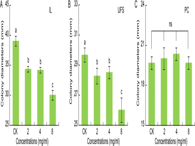
The crude extract was added to the cooled PDA medium at working concentrations of 0 (CK), 2, 4, and 8 mg/ml. Mycelial plugs (4-mm diameter) of the fungi (IL and UFS), isolated from H. cordata, were cut from the leading edge of colonies on PDA and inoculated into the center of PDA plates, which were augmented with the appropriate working concentration of the crude H. cordata rhizome extract. Considering that upon transfer of the plugs of PC, the spores may fall off and form several new colonies on the PDA, which would affect the experimental results, spores of PC (isolated from H. cordata) were transferred with a dissecting needle instead of plugs. The culture conditions were 28°C in the dark. Colony diameter (CD) was measured after eight days, and the experiment was repeated three times.
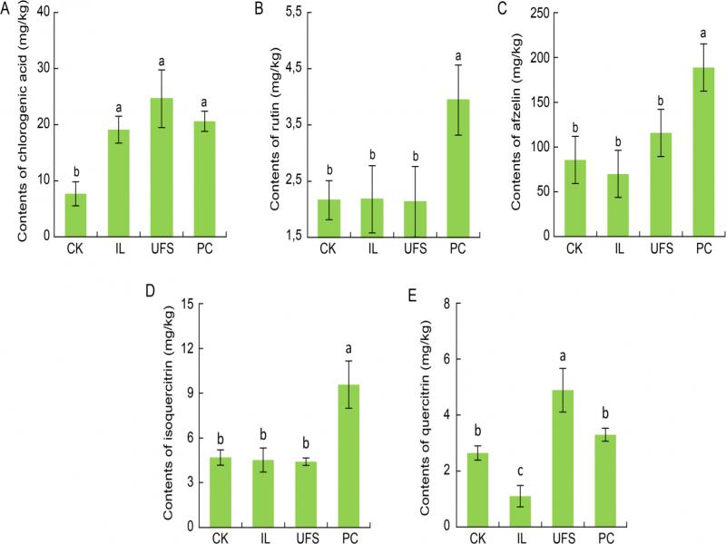
HPLC analysis of phenolics. The phenolic compounds were extracted from the rhizomes (the sterile H. cordata seedlings grown for 12 days in non-sterile soil inoculated with LPFs and CK), and analyzed using high-performance liquid chromatography (HPLC). The phenolics were extracted from H. cordata rhizomes (fresh weight) from each treatment; the fresh leaves (≈ 0.1 g) pulverized using a homogenizer were placed into a 20-ml glass storage bottle (Xianglong, China), with a tight-fitting lid and mixed with methanol (5 ml). The bottle was then sealed and subjected to ultrasonic extraction for 30 min. The extracted solutions were centrifuged and filtered through a 0.45-μm-nylon membrane filter (Jinlong, China). Each sample extract was stored at 4°C in the dark until analysis was carried out. Stock solutions (10 µg/ml in methanol) of phenolic standards were prepared, namely the phenolic acid, chlorogenic acid, flavonoid quercetin, the flavonoid glycosides rutin, isoquercitrin, quercitrin, and afzelin.
The concentrations of these phenolics in H. cordata crude rhizome extracts was determined using a slightly modified version of the method reported by Yang et al. (2014). The conditions included an HPLC instrument (LC-20AT; Shimadzu, Japan), a SHIM-PACK C18 CLC ODS reversed-phase chromatographic column (150 × 6.0 mm i.d.), with detection at 345 nm wavelength and a column temperature of 40°C. The linear solvent gradient, consisting of solvent A (acetonitrile: methanol = 11 : 5 (v/v)) and solvent B (0.1% formic acid (v/v), was conducted for sample elution as follows: 6% A (0 ~ 1 min); 6 ~ 13.2% A (1 ~ 6 min); 13.2 ~ 18.0% A (6 ~ 9 min); 18.0 ~ 18.9% A (9 ~ 11 min); 18.9 ~ 29.1% A (11 ~ 31 min); 29.1 ~ 37.9% A (31 ~ 48 min); 37.91 ~ 100% A (48 ~ 55 min); 100% A (55 ~ 65 min); 1001 ~ 6% A (65 ~ 66 min); and 6% A (66 ~ 75 min). The flow rate program was conducted as follows: 1.2 ml/min (0–1 min); 1.2–1.39 ml/min (1 ~ 6 min); 1.39 ~ 1.10 ml/min (6 ~ 9 min); 1.10 ~ 1.0 ml/min (9 ~ 11 min); 1.0 ~ 1.10 ml/min (11 ~ 31 min); 1.10 ~ 1.20 ml/min (31 ~ 48 min); 1.20 ~ 1.39 ml/min (48 ~ 55 min); and 1.20 ml/min (55 ~ 75 min). Working solutions of the standards, chlorogenic acid, rutin, isoquercitrin, quercitrin, afzelin, and quercetin were obtained by dilution of the individual standard solutions and the mixed-standard stock solutions of the six phenolics with methanol. The working solutions were analyzed by HPLC. The concentrations of each of the six phenolics in the H. cordata were calculated based on the peak areas of the individual compounds. The concentration of the individual phenolics was calculated by the external standard method, based on the peak areas from a HPLC chromatographic file of phenolics.
Statistical analysis. Microsoft Office Excel 2010 was used to analyze the raw data for every treatment, with every parameter measured in three replicate samples. Summary statistics were mean ± standard deviation (SD). Data analysis was carried out by one-way analysis of variance (ANOVA) following Tukey posthoc analysis using the R programming language.
Results
Isolation and identification of latent pathogens from H. cordata rhizomes. Our aim was to investigate how the latent pathogens were present but constrained stably in the healthy H. cordata rhizomes. We found that the rhizomes were heavily colonized when the healthy rhizomes, following surface sterilization, were cultured on a half-strength MS medium (Fig. S1). We isolated the LPFs, which emerged from inside the surface-sterilized H. cordata rhizomes on PDA. Fig. 2 and Table SI showed that three fungal species isolated were identified as I. liriodendra (IL), an unidentified fungal sp. (UFS), and P. citrinum (PC), using ITS1-5.8S-ITS2. They were numbered NSF-1, NSF-2, and NSF-3, respectively (Fig. 1 and Table SII). IL proliferated rapidly on the PDA medium. The surface mycelium was pure white with edges, with abundant aerial mycelium, shaped like cotton wool. The hyphae had few branches; colonies were light yellow to dark red on the reverse side of the plate three-septate conidia were long and cylindrical (Fig. 3A-C). UFS colonies had a gray woolly appearance, with white hyphae on the outside and edges, gray to dark black on the reverse side, and obvious concentric annular bands. Hyphae were slender and loose, with few or no branches; two forms of hyphae coexisted, one with a smooth surface and the other with dense cup-shaped protrusions, and no spores were observed (Fig. 3D-F). Colonies of PC had a gray green felt-like appearance with white edges. After about two weeks of growth, the colony gradually stopped extending and formed white, velvety, aerial hyphae on the original colony, white to yellow on the reverse. The hyphae were dense and separate, the sporangia were broom-like, the conidia were light green and round, and were easy to spread (Fig. 3G-I).
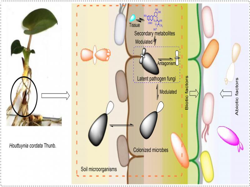
Fig. 1.
The hypothesis of maintaining the stable colonized microbes of the plant-LPFs in H. cordata.
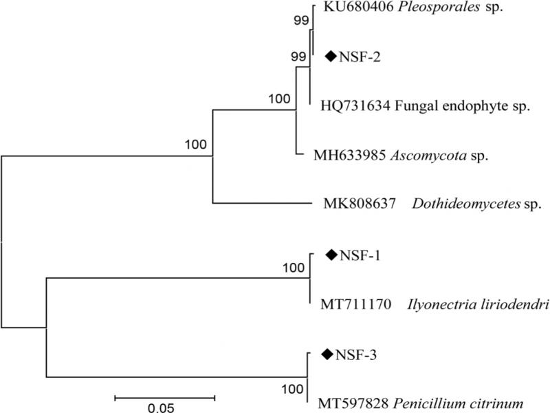
Fig. 2.
Phylogenetic tree of the fungi isolated from the rhizomes of H. cordata based on sequences of the ITS region.
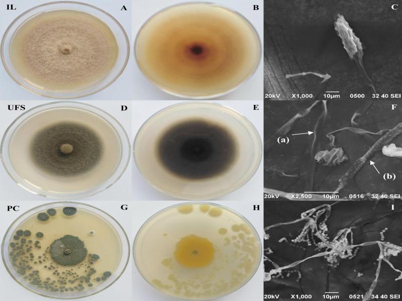
Fig. 3.
LPFs isolated from stems of H. cordata: A) surface side, B) reverse side and C) a germinated spore of IL on PDA, D) surface side, E) reverse side and F) two types including smooth (a) and rough (b) of mycelium of UFS on PDA, G) surface side, H) reverse side and I) broom-shaped sporangia of PC on PDA. IL – I. liriodendri, UFS – an unidentified fungal sp., PC – P. citrinum.
Growth properties of H. cordata-colonized LPFs in sterile soils and non-sterile soils. To investigate the interactions between the colonizing microbiota and the LPFs within the rhizomes, we investigated any possible interactions between the colonizing microbiota (originating from nonsterile soils) and the LPFs. To achieve this goal, we investigated whether the growth of the sterile H. cordata seedlings in the nonsterile soils (containing soil microorganisms) and the sterile soils were affected by IL, UFS, and PC (Fig. 4 and 5). When the sterile H. cordata seedlings growing in sterile soils were inoculated with IL, the growth was strongly negatively affected (Fig. 4B). Compared with CK (Fig. 4A) (the sterile water was added to the sterile H. cordata seedlings growing in sterile soils), the control seedling significantly grew faster than the inoculated seedlings. The color of the young leaves of the seedlings began to fade on the 3rd day, after which the whole plant quickly wilted, and, by 15 days, the plants were completely dead, and dead tissue showed obvious white mycelium.
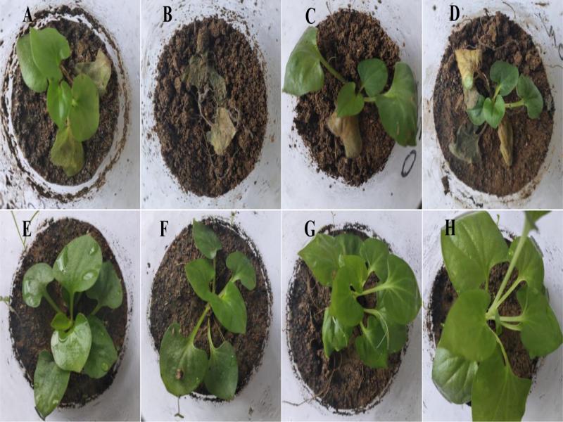
Fig. 4.
Direct inoculation assays showed the effects of the LPFs on H. cordata sterile seedlings in sterilized soils (A, B, C and D) and non-sterile soils (D, E, F and H) at the 15th day.
A) the sterile seedling was watered with sterile water as a control in sterilized soils, B) the sterile seedling was added with I. liriodendri in sterilized soils, C) the sterile seedling was added with an unidentified fungal sp. in sterilized soils, D) the sterile seedling was added with P. citrinum in sterilized soils, E) the sterile seedling was watered with sterile water as a control in non-sterile soils, F) the sterile seedling was added with I. liriodendri in non-sterile soils, G) the sterile seedling was added with an unidentified fungal sp. in non-sterile soils, H) the sterile seedling was added with P. citrinum in non-sterile soils.
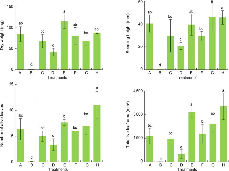
Fig. 5.
The effects of inoculating the LPFs in sterilized soils (A, B, C and D) and non-sterile soils (D, E, F and H) on H. cordata at the 15th day.
A) the sterile seedling was watered with sterile water as a control in sterilized soils, B) the sterile seedling was added with I. liriodendri in sterilized soils, it died, C) the sterile seedling was added with an unidentified fungal sp. in sterilized soils, D) the sterile seedling was added with P. citrinum in sterilized soils, E) the sterile seedling was watered with sterile water as a control in non-sterile soils, F) the sterile seedling was added with I. liriodendri in non-sterile soils, G) the sterile seedling was added with an unidentified fungal sp. in non-sterile soils, H) the sterile seedling was added with P. citrinum in non-sterile soils. Different letters in the one group represent significant differences (p < 0.05).
When the sterile H. cordata seedlings growing in sterile soil were inoculated with UFS, the effects on H. cordata seedling growth were not significantly adverse. However, the average of dry weight, seedling height, number of live leaves, and total leaf area declined when compared with CK (Fig. 4C and 5). When the sterile H. cordata seedlings growing in sterile soil were inoculated with PC (Fig. 4D), the old leaves began to fade after five days. The plant growth slowed down, compared with CK (Fig. 4A), and after 15 d, the old leaves had died, but the young leaves remained green, the dry weight, seedling height, and total leaf area declined by 50.5%, 48.6, and 69.8%, respectively (p < 0.05) (Fig. 5). When the sterile H. cordata seedlings grown in non-sterile soil were inoculated with IL, UFS, or UFS, and compared to CK (the sterile H. cordata seedlings grown in non-sterile soils and treated with sterile water) (Fig. 4E), the growth of H. cordata seedlings was barely affected (Fig. 4F-H). Furthermore, the growth of sterile H. cordata seedlings in sterile soils (Fig. 4A) was significantly slower than the growth in non-sterile soils (Fig. 4E); the total live leaf area increased 47.9% (p < 0.05) (Fig. 5).
Antagonism between LPFs. The LPFs isolated from the rhizomes colonized the healthy H. cordata rhizome (Fig. S1) with no symptoms, suggesting that they formed a stable system in which the pathogenic factors of LPFs were suppressed. The results from our in vitro studies showed that the colony diameters of the three strains (IL, UFS and PC) were significantly inhibited when three strains were grown in pairs for seven days in the antagonism assay on the PDA medium (Fig. 6). It was clear that, after seven days, the mycelia of the pairs of LPFs in the antagonism trials were not in obvious contact. IL significantly inhibited the colony growth of UFS, with an inhibition rate (IR) of 85.7% (Fig. 6A and Table I), and PC (IR = 52.1%, Fig. 6A and Table I). UFS significantly inhibited colony growth of IL (IR = 26.1%, Fig. 6B and Table I), and PC (IR = 18.5%, Fig. 6C and Table I). PC significantly inhibited colony growth of IL (IR = 52.6%, Fig. 6A and Table I), and UFS (IR = 62.2%, Fig. 6C and Table I). These findings confirm that there is a competitive relationship between any pair of the LPFs.
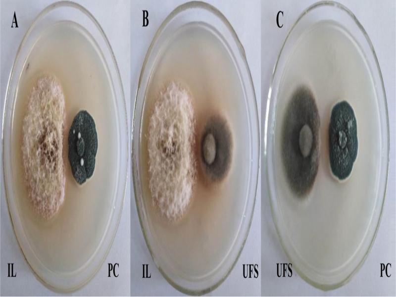
Fig. 6.
A direct antagonisms on PDA medium. Antagonism assays among LPFs: 4 mm fungal plugs were placed 3 cm at distance and cultured at 28°C in darkness. The image was taken at the 7th day.
A) interaction between IL and PC, B) interactions between IL and UFS, C) interactions between UFS and PC. IL – I. liriodendri, UFS – an unidentified fungal sp., PC – P. citrinum.
Table I
Colony radius (CR) and inhibition rate (IR) of mycelia grown and the colony radius compared with other fungi on PDA at the 7th day.
| Strains | CK | IL | UFS | PC | |||
|---|---|---|---|---|---|---|---|
| CR (mm) | CR (mm) | IR (%) | CR (mm) | IR (%) | CR (mm) | IR (%) | |
| NSF-1 | 27.2 ± 0.5a | – | – | 21 ± 0.8b | 26.7 | 15.0 ± 0c | 52.6 |
| NSF-2 | 22.2 ± 0.5a | 6.7 ± 1.5b | 85.7 | – | – | 10.7 ± 1.2c | 62.2 |
| NSF-3 | 15.9 ± 0.4a | 9.7 ± 0.6b | 52.1 | 13.7 ± 1.2c | 18.5 | – | – |
| Concentrations (mg/ml) | IL IR (%) | UFS IR (%) | PC IR (%) |
|---|---|---|---|
| 0 | – | – | – |
| 2 | 11. 97 | 4.49 | -1.69 |
| 4 | 12.39 | 3.37 | -3.39 |
| 8 | 23.08 | 12.36 | 0.00 |