
Table I
Description of 82 participants of this study.
| Group | Vaginal delivery (n = 51) | Cesarean delivery (n = 31) | p-value |
|---|---|---|---|
| Sex (%) | 0.907 | ||
| Boys | 24 (47.1%) | 15 (48.4%) | |
| Girls | 27 (52.9%) | 16 (51.6%) | |
| Gestational age (w) | 39.0 ± 0.9 | 39.2 ± 1.0 | 0.532 |
| Birth weight (g) | 3,091.0 ± 299.7 | 3,117.4 ± 260.4 | 0.685 |
| Hb (g/l) | 163.9 ± 12.0 | 161.2 ± 10.2 | 0.296 |
| Premature rupture of membranes (h) | 3.2 ± 2.1 | 3.3 ± 2.5 | 0.836 |
| Feeding (%) 3-day-old | 0.687 | ||
| Breast-fed | 24 (47.1%) | 12 (38.7%) | |
| Mixed-fed | 16 (31.4%) | 10 (32.3%) | |
| Fomula-fed | 11 (21.6%) | 9 (29.0%) | |
| Feeding (%) 30-42-day-old | 0.853 | ||
| Breast-fed | 21 (41.2%) | 11 (35.5%) | |
| Mixed-fed | 16 (31.3%) | 10 (32.3%) | |
| Fomula-fed | 14 (27.5%) | 10 (32.3%) | |
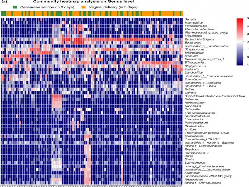
Fig. 1.
Intestinal microbiota community structure of 2 groups (vaginal delivery and cesarean section) in 3 days neonates.
(A) Intestinal microbiota community heatmap analysis on the genus level of the two groups.
(B) Wilcoxon rank-sum test bar plot on the genus level between the two groups.
(C) Principal coordinate analysis (PCoA) on unweighted UniFrac distances between the neonatal microbiota is shown along the first two principal coordinates (PC) axes. Each point represents a single sample and is colored by a delivery mode: vaginal delivery, blue; cesarean section, red. The closer the two sample points are, the more similar the species composition is.
(D) PCoA box diagram. Represents the discrete distribution of different groups of samples on the PC1 axis: vaginal delivery, blue; cesarean section, red.
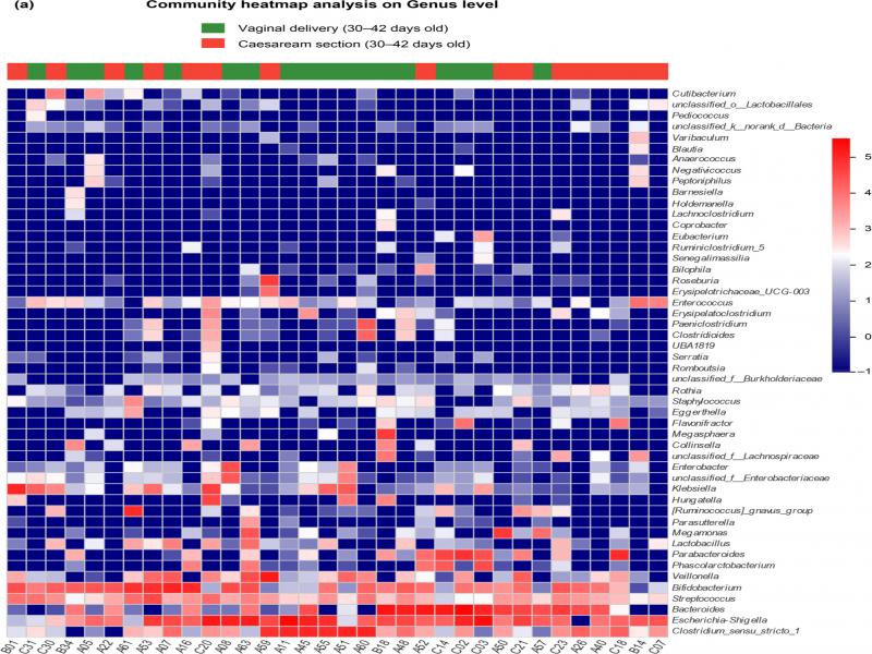
Fig. 2.
Intestinal microbiota community structures of two groups (vaginal delivery and cesarean section) in 30–42 days infants.
(A) Intestinal microbiota community heatmap analysis on the genus level of the two groups.
(B) Wilcoxon rank-sum test bar plot on the genus level between the two groups.
(C) Principal coordinate analysis (PCoA) on unweighted UniFrac distances between the infants’ intestinal microbiota is shown along the first two principal coordinates (PC) axes. Each point represents a single sample and is colored by delivery mode: vaginal delivery, blue; cesarean section, red. The closer the two sample points are, the more similar the species composition is.
(D) PCoA box diagram. Represents the discrete distribution of different groups of samples on the PC1 axis: vaginal delivery, red; cesarean section, blue.
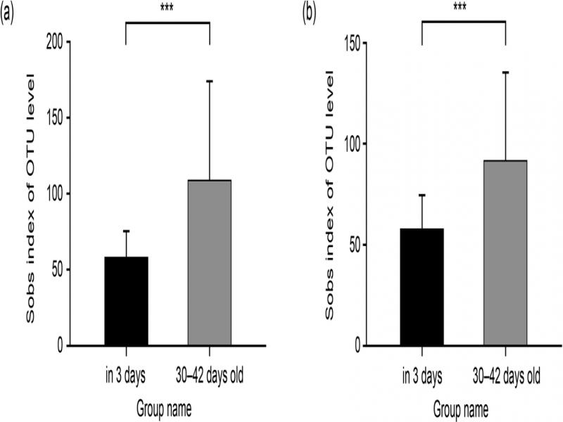
Fig. 3.
Student’s t-test for sobs index of vaginal delivery and cesarean section groups’ intestinal microbiota at two points (in 3 days and 30–42 days old). (A) Sobs index at the two points of vaginal delivery infants. (B) Sobs index at the two points of cesarean section infants.
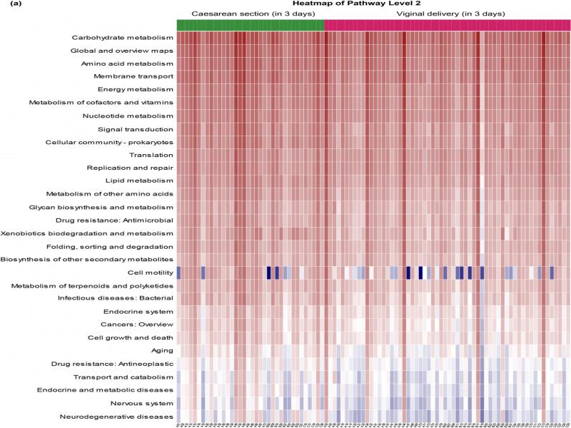
Fig. 4.
Predicted function: Heatmap of KEGG pathway level 2.
(A) Heatmap showing distinct microbial gene (KEGG pathway level 2) profiles of the two groups’ stool in 3 days after delivery: vaginal delivery, r)ed; cesarean section, green.
(B) Heatmap showing distinct microbial gene (KEGG pathway level 2) profiles of 2 groups in 30–42 days old: vaginal delivery, green; cesarean section, red.