
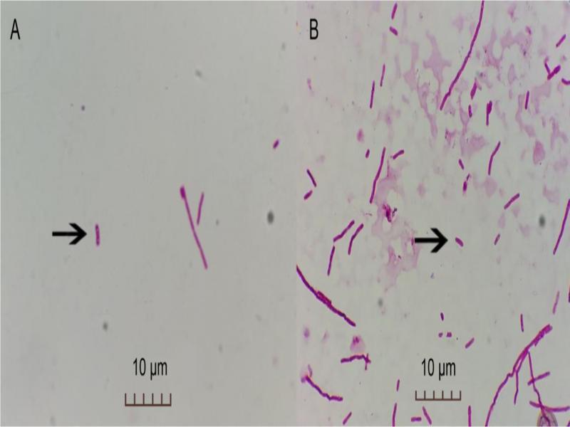
Fig. 1.
The Gram staining results of E. coli. A – Capillary sample, B – Glass slide sample.
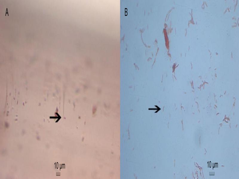
Fig. 2.
The Gram staining results of V. parahaemolyticus. A – Capillary sample, B – Glass slide sample.
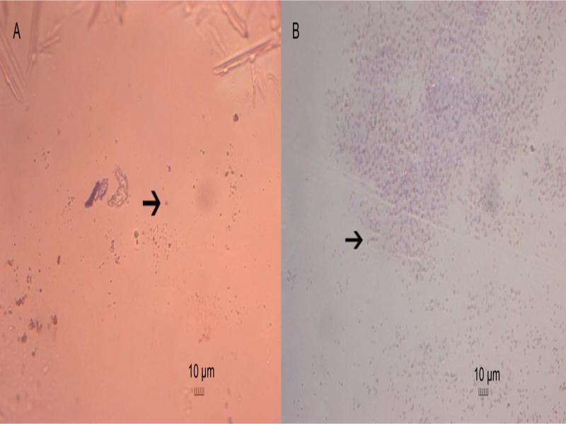
Fig. 3.
The Gram staining results of S. marcescens. A – Capillary sample, B – Glass slide sample.
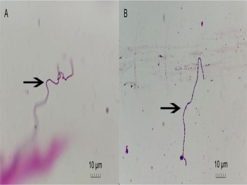
Fig. 4.
The Gram staining results of S. thermophiles. A – Capillary sample, B – Glass slide sample.
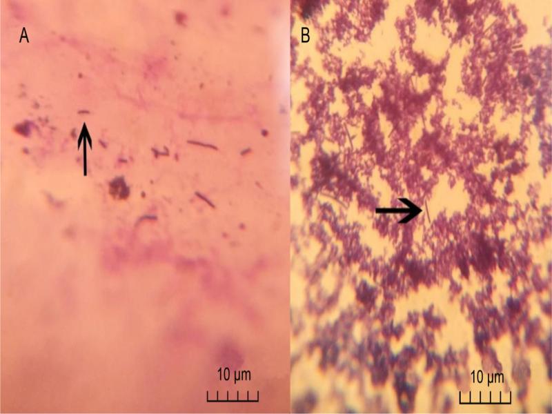
Fig. 5.
The Gram staining results of L. bulgaricus. A – Capillary sample, B – Glass slide sample.
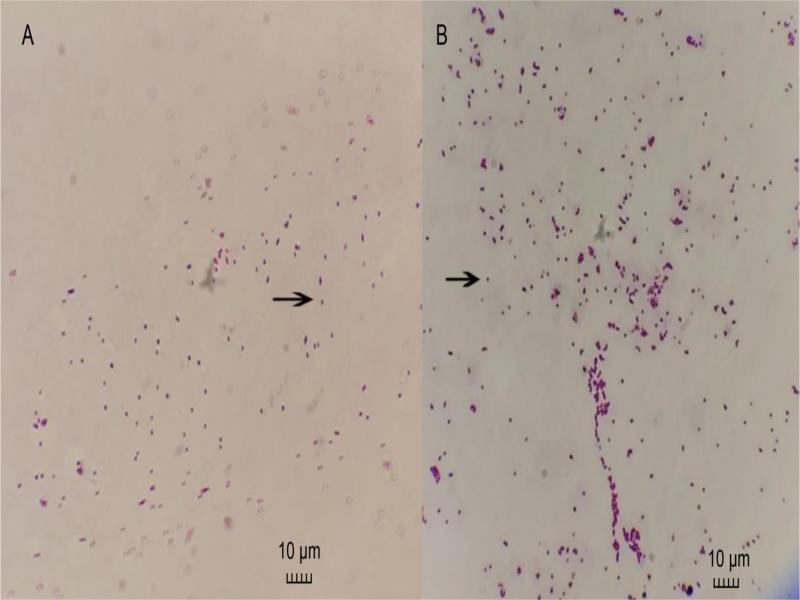
Fig. 6.
The Gram staining results of S. aureus. A – Capillary sample, B – Glass slide sample.
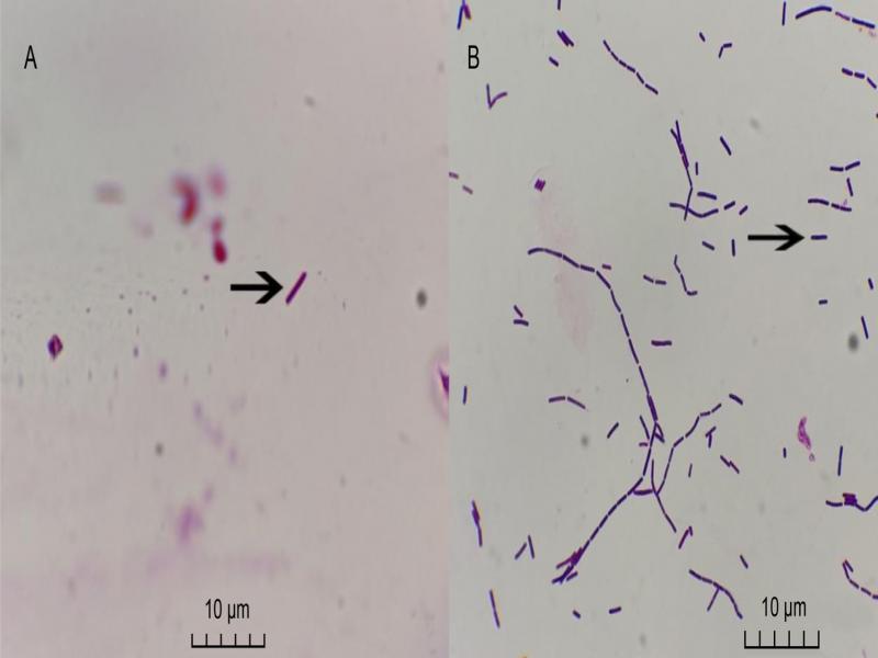
Fig. 7.
The Gram staining results of B. subtilis. A – Capillary sample, B – Glass slide sample.
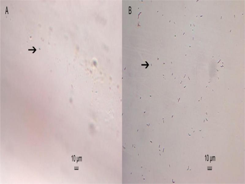
Fig. 8.
The Gram staining results of B. Licheniformis. A – Capillary sample, B – Glass slide sample.
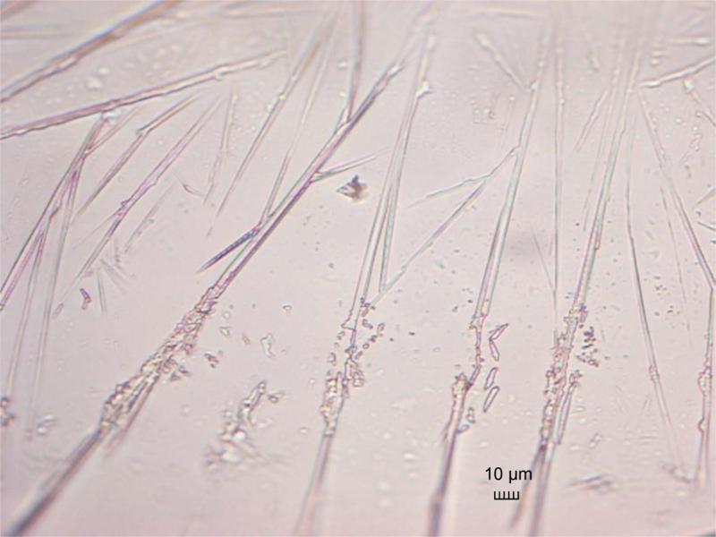
Fig. 9.
The microstructure of the overheated capillary tube.