
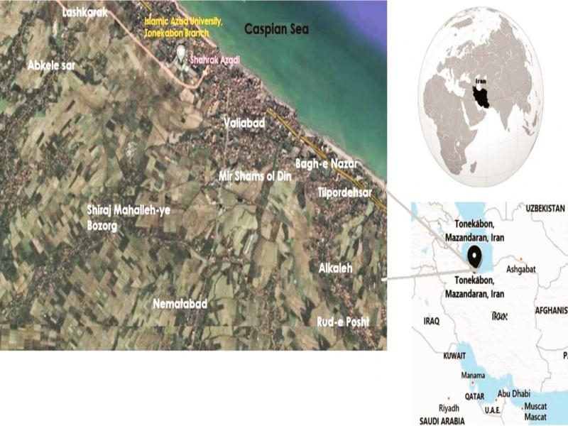
Fig. 1.
Localization of ten environmental sites investigated.
Table I
The specific primers of Leptospira.
| Name of primer | Type | Sequence of primer |
|---|---|---|
| 16S rRNA | Forward | 5’-GAACTGAGACACGGTCCAT-3’ |
| 16S rRNA | Reverse | 5’-GCCTCAGCGTCAGTTTTAGG-3’ |
| LipL32 | Forward | 5’-ATCTCCGTTGCACTCTTTGC-3’ |
| LipL32 | Reverse | 5’-ACCATCATCATCATCGTCCA-3’ |
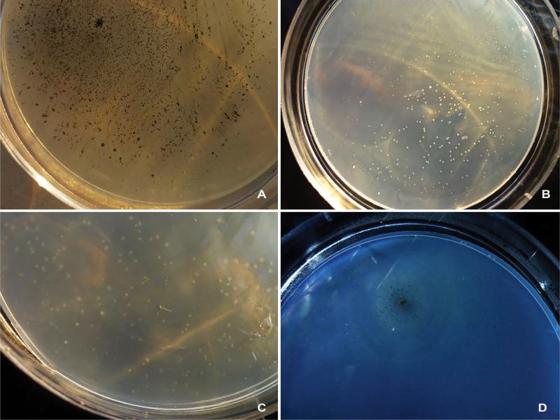
Fig. 2.
Leptospira colonies.
(a) Formation of surface’s black colonies, with round shape, needle tip size (at 30 days of incubation on EMJH agar), wooly margins, and hilly elevation related to Leptospira pathogen species. (b) Formation of sub surface’s white colonies with round shape, small size, and some smaller colonies, size 1–2 mm (after 30 days of incubation on EMJH agar), smooth (entire) margins, flat elevation related to Leptospira perdikensis. (c) Formation of sub surface’s very bright cream colonies with round shape, small size, 1–3 mm, (after 30 days of incubation on EMJH agar), smooth (entire) margins, with small to medium halo, flat elevation related to Leptospira ryugenii. (d) It had the characteristics of a colony (a), except that in this case, rings were seen to the same distance around the surface black colony related to Leptospira pathogen species.
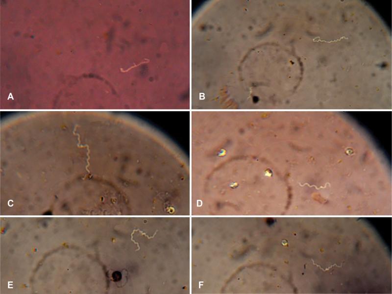
Fig. 3.
Microscopic observation of Leptospira with a 100× magnification optical microscope
A: Multiplying Leptospira; the image relates to pathogenic Leptospira spp., B: The Leptospira bacterium that is twisted over itself is located on the right. In the lower-left a shorter Leptospira is visible; the image related to Leptospira ryugenii, C and E: Long Leptospira, D: The hook area of the Leptospira bacterium was clearly observed in a microscopic view; the image related to Leptospira perdikensis, F: Two Leptospira bacteria that are interconnected from one end and form a long separable leptospiral chain; the image related to Leptospira sp.
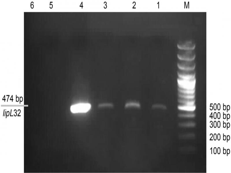
Fig. 4.
Agarose gel electrophoresis (1.5%) of PCR products of environmental samples.
Columns 1, 2, and 3, pathogenic Leptospira spp.; Column 4 – a reference strain; Column 5 – a negative control; Column 6 – E. coli; Column M – 100 bp DNA ladder.
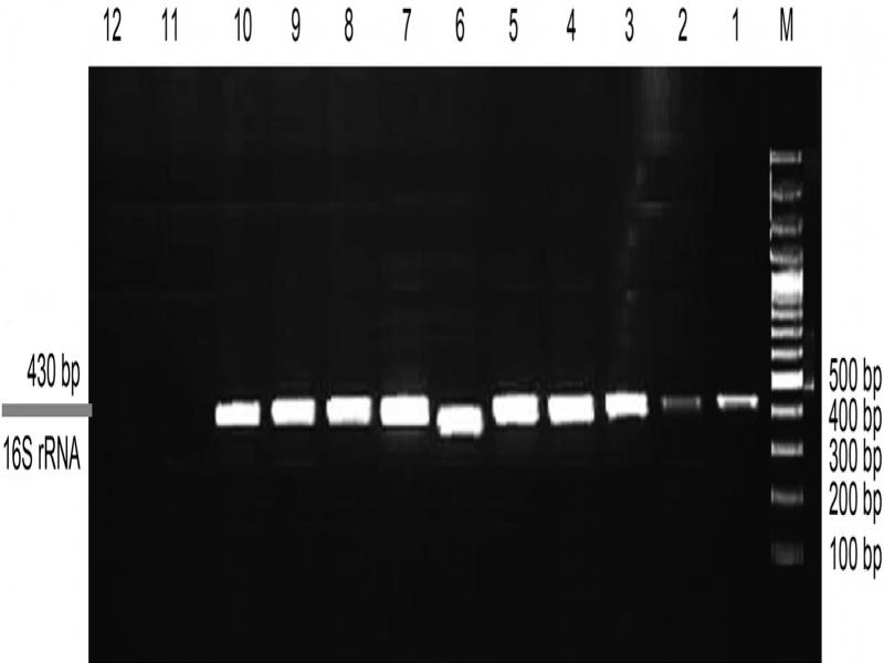
Fig. 5.
Agarose gel (1.5%) analysis of a PCR products of environmental samples; Columns 1–9 – species of Leptospira; Column 10 – a reference strain; column 11 – a negative control; Column 12 – E. coli E. coli; Column M – 100 bp DNA ladder. Sampling sites that were positive for the presence of pathogenic Leptospira by both culture and PCR were reported in Fig. 1 and Table II.
Table II
The sampling sites and positive cases reported in the study.
| Number of positive cases of saprophytic Leptospira | Number of positive cases of pathogenic Leptospira | Number of samples | Site |
|---|---|---|---|
| 3 | 2 | 5 | Valiabad |
| 2 | 2 | 5 | Bagh e nazar |
| 0 | 1 | 5 | Mir shams-ol |
| 1 | 1 | 5 | Rud e posht |
| 1 | 0 | 5 | Alkaleh |
| 1 | 0 | 5 | Abkele sar |
| 0 | 1 | 5 | Lashkarak |
| 1 | 1 | 5 | Nematabad |
| 1 | 1 | 5 | Shiraj mahalleh-ye bozorg |
| 1 | 1 | 5 | Tilpordehsar |
| 11 (22%) | 10 (20%) | 50 | Total |
Table III
The results of the sequencing.
| Molecular detection | Max Score | Total Score | Query Cover | E Value | Per Ident | Accession |
|---|---|---|---|---|---|---|
| Leptospira perdikensis strain CES 16S ribosomal RNA gene | 612 | 612 | 93% | 3e-171 | 99.41% | MN086353.1 |
| Leptospira ryugenii strain CES 16S ribosomal RNA gene | 176 | 176 | 26% | 5e-40 | 100% | MN086356.1 |
| Leptospira sp. MS336 gene for 16S ribosomal RNA gene | 593 | 593 | 100% | 2e-166 | 100% | AB758752.1 |
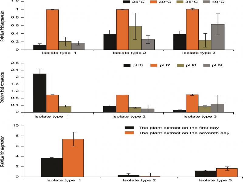
Fig. 6.
Comparison of the lipL32 gene expression in pathogenic Leptospira under the influence of temperature, pH, and Ph. amarus plant extract.