Introduction
In the last decade, there has been a marked increase in the frequency of breast implantation in patients undergoing surgical treatment for breast cancer. The percentage of patients having breast reconstruction after a mastectomy is as high as 36.4–43.3% (NCIN 2011; Ilonzo et al. 2017). However, this applies to data from specialized treatment centers for patients with breast cancer. In other facilities, the percentage does not usually exceed 20% of the cases of mastectomy (Alderman et al. 2006). The frequency of such treatments in Poland is much lower. However, there is a lack of accurate data on the above problem on a national scale. The previous studies, which are yet not numerous, have shown that a percentage of the patients undergoing mastectomy and breast reconstruction may reach 22.4% (Tarkowski et al. 2017). However, the complications that accompany the introduction of the implant (expander, final prosthesis) remain a major challenge for oncological surgeons. The most frequent complications include infections; it is estimated that up to 29% (mean 5.8%) of breast reconstruction surgery is complicated by infection (Phillips et al. 2013). Infection is cost-intensive and 70–80% of patients ultimately require removal of the implant (Pittet et al. 2005; Seng et al. 2015).
Among the risk factors for infection are associated diseases such as diabetes, renal failure, and skin diseases, but also systemic treatment and radiotherapy. Other factors that increase the risk of infection include obesity and nicotinism. Infections are also triggered by factors associated with the surgery itself: an immediate breast reconstruction with the use of a final prosthesis is more often complicated by infection than deferred reconstruction. The prolonged (> 2 h) duration of surgery and post-operative drainage also have an unfavorable effect (Pittet et al. 2005; Araco et al. 2007).
Among the etiological agents of infections, the most common are skin microbiota: coagulase-negative Staphylococcus, Corynebacterium spp., Propioni-bacterium acnes, and Staphylococcus aureus, but more and more frequently there are reports on the increased proportion of Gram-negative bacteria from the order Enterobacterales and anaerobic microorganisms among etiological agents of these infections (Halvorson et al. 2007; Weichman et al. 2013; Seng et al. 2015).
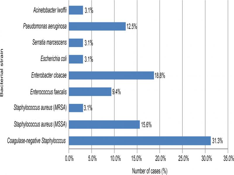
This study aimed to retrospectively analyze the causes of removal of implants after mastectomy in the course of breast cancer treatment and to determine the frequency and time of appearance of implant infections as well as the etiological agents causing them.
Experimental
Materials and Methods
The study involved 428 patients treated in one oncological center in the years 1998–2018, who underwent a total of 648 breast reconstruction procedures using artificial implants.
In each case, the implantation procedure was preceded by the test for MRSA (Methicillin-Resistant Staphylococcus aureus) and MSSA (Methicillin-Sensitive Staphylococcus aureus). The swabs taken for this purpose came from the nasal vestibule, palms, and axilla on the side of the primary tumor of the operated patients. The materials for microbiological tests were seeded on Columbia Agar supplemented with nalidixic acid and amikacin + 5% sheep blood (bioMérieux, France) as well as chromogenic Brilliance MRSA (Oxoid, UK). After 24 h, the morphology of the colonies was assessed, and a catalase and coagulase assays were performed. Also, the sensitivity to methicillin was assessed following the current recommendations of the National Reference Center for Antimicrobial Susceptibility using the disk diffusion method.
Mupirocin eradication was implemented in cases of MRSA colonization in the nasal vestibule. If screening did not show the presence of MRSA, eradication was not performed. On the day before the treatment, in the evening hours, a whole-body cleansing was recommended with the use of an antiseptic intended for skin decontamination or with the body sponges impregnated with an antiseptic (chlorhexidine 4% soap solution). On the day of treatment, the whole-body cleansing was again recommended with the use of an antiseptic intended for skin decontamination. The preparation of the patient for a surgery proceeded under the standards of nursing practice currently in force at the hospital.
In all patients, perioperative prophylaxis was routinely used (cefazolin in a dose of 1.0 g in an intravenous injection – given every 8 hours for 5 days). The first dose of antibiotics was given up to 30 min before the surgery. If the drain remained longer than 5 days, antibiotic therapy was prolonged until the drain was removed. The procedure for perioperative antibiotic prophylaxis was in line with the procedure adopted in our center, i.e., the prolonged antibiotic prophylaxis in the case of wound drainage. Notwithstanding the currently recommended use of 1 dose of antibiotic (Phillips et al. 2013), the procedure referred to the results of the research presented by (Brand et al. 1993), which showed greater effectiveness of longer-term use of antibacterial drugs in the prophylaxis of infectious complications after implantation.
From the group of 428 patients with artificial implants used for breast reconstruction, 44 patients were selected for the analysis when it was necessary to remove the implant, which accounted for 10.3% of the patients that underwent surgery. In two out of the 44 patients (4.5%), this occurred twice (after twice reconstructed breast, with a time interval between subsequent surgical procedures – 26 and 27 months, respectively), in one patient (2.3%), the above situation concerned the reconstruction of both breasts (carried out on two different dates – with an interval of 20 months). In none of these three patients were signs of infection of the surgical site before the second surgery. Thus, the total number of removals of implants concerned 47 cases. This accounted for 7.3% of all surgical procedures associated with generative treatment after implantation of an artificial breast implant. The incidence of infection was found in 39 cases, which accounted for 6.0% of all reconstructive procedures. Further epidemiological analyses were based on the number of the performed procedures, which resulted in the loss of the implant.
In 20 cases (42.6%), the need to remove the implant concerned immediate breast reconstruction surgery, while in the remaining 27 cases (57.4%), deferred reconstruction had been performed. The type of reconstructive implant used during the procedure (expander, expander prosthesis, or final prosthesis) as well as the duration of the procedure (immediate or deferred reconstruction) was the result of the current organizational arrangements for treatment and financing of the surgical procedures. They were not the result of the planned choice of the patients.
In 46 cases, the reconstruction of the amputated breast gland consisted of the insertion of an implant in the large pocket created behind the pectoral muscle. In one case, the patient’s tissues were used to cover the implant (pedunculated skin-muscle flap taken from the latissimus dorsi muscle).
In 13 cases (27.7%), the reconstruction was preceded by chemotherapy and radiotherapy, in one case (2.1%) – by radiotherapy (this was the case in most patients who underwent restorative treatment in the deferred mode). In 13 cases (27.7%) included in the study, the patients required pre-operative chemotherapy (regardless of the mode of reconstruction). In the remaining patients, the surgery was not preceded by any other type of anticancer treatment (in most cases, these were immediate reconstruction treatments), as it is presented in Table I.
Table 1.
Characteristics of pre- and post-implantation treatment of cancer patients.
| Characteristic | Number of cases n = 47 n (%) |
|---|---|
| Pre-implantation treatment: | |
| – RTH | 1 (2.1) |
| – CHTH | 13 (27.7) |
| – RTH+CHTH | 13 (27.7) |
| – No treatment | 19 (40.4) |
| – No data | 1 (2.1) |
| Post-implantation treatment: | |
| – RTH | 1 (2.1) |
| – CHTH | 4(8.5) |
| – RTH+CHTH | 1 (2.1) |
| – No treatment | 40 (85.1) |
| – No data | 1 (2.1) |
| Characteristics | Number of cases n = 47 n (%) |
|---|---|
| Type of carcinoma | |
| Ductal carcinoma | 36 (76.6) |
| Lobular carcinoma | 2 (4.3) |
| Other forms of invasive cancer | 3 (6.4) |
| DCIS | 4 (8.5) |
| No data | 2 (4.3) |
| Clinical stage (cTNM) | |
| IA | 14 (29.8) |
| IIA | 17 (36.2) |
| IIB | 5 (10.6) |
| IIIA | 2 (4.3) |
| IIIB | 1 (2.1) |
| No data | 8 (17.0) |
| Diabetes | |
| Yes | 3 (6.4) |
| No | 44 (93.6) |
| Nicotinism | |
| Yes | 2 (4.3) |
| No | 36 (76.6) |
| No data | 9 (19.1) |
| BMI [kg/m2] | |
| < 25 | 21 (44.7) |
| ≥ 25 | 26 (55.3) |
| MSSA carrier | |
| Yes | 8 (17.0) |
| No | 26 (55.3) |
| No data | 13 (27.7) |
| Type of reconstruction | |
| Immediate | 20 (42.6) |
| Deferred | 27 (57.4) |
| Type of implant | |
| Expander | 23 (48.9) |
| Expander prosthesis | 8 (17.0) |
| Prosthesis | 5 (10.6) |
| Expander prosthesis/prosthesis (2nd stage of reconstruction - replacement of implant after earlier implantation of expander) | 11 (23.4) |
| Characteristics | Number of cases n = 47 n (%) |
|---|---|
| Infection | |
| Early (≤ 30 days) | 6 (12.8) |
| Late (> 30 days) | |
| 31-90 days | 11 (23.4) |
| 91-365 days | 17 (36.2) |
| > 365 days | 5 (10.6) |
| – Local recurrence of breast cancer | 2 (4.3) |
| – Local recurrence of breast cancer in the chest wall | 2 (4.3) |
| – Postoperative pain | 1 (2.1) |
| – Leakage of prosthesis/expander prosthesis | 2 (4.3) |
| – No data | 1 (2.1) |