
Table I
Strains and plasmids used in this study.
| Strains/Plasmids | Description | Source |
|---|---|---|
| Strains | ||
| Streptococcus iniae HNM-1 | Isolated from infected A. sinensis | This study |
| Escherichia coli DH5α | Used for cloning and propagation of plasmids | Novagen |
| Escherichia coli BL21(DE3) | Used for protein expression | Invitrogen |
| Salmonella typhimurium | This study | |
| Staphylococcus aureus | This study | |
| Acinetobacter baumannii | This study | |
| Pseudomonas aeruginosa | This study | |
| Plasmids | ||
| pMD19-T | Carries ampR gene; used for cloning PCR product with A at 3’ ends | Takara |
| pET 22b (+) | Carries ampR gene; used for expressing S. iniae Alanine racemase | Novagen |
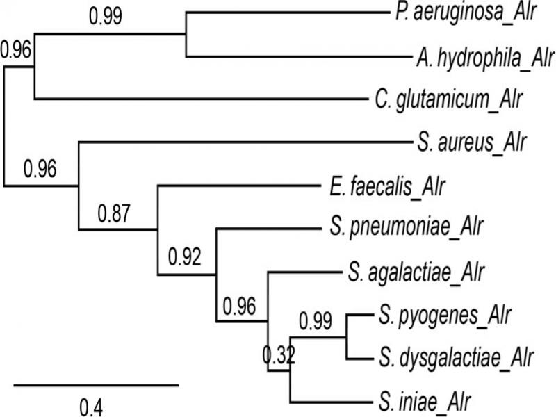
Fig. 1.
Phylogenetic relationships of SiAlr and homologs alanine racemases from Pseudomonas aeruginosa PAO1, Corynebacterium glutamicum ATCC 13032, Aeromonas hydrophyla ATCC 7966, Staphylococcus aureus ABFQT, Enterococcus feacalis D32, Streptococcus pneumoniae MDRSPN001, Streptococcus agalactiae 2603V/R, Streptococcus pyogenes MGAS10750 and Streptococcus dysgalactiae Kdys0611. The tree was constructed using Neighbor end-joining. Maximum likelihood tree based on complete coding sequences deposited in Genbank. The evolutionary distances were computed using the p-distance method and are reported in the units of the number of amino acid differences per site.
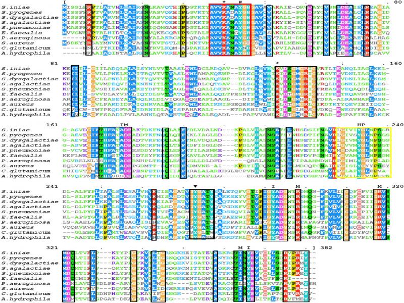
Fig. 2.
Structure-based sequence alignment of alanine racemases sequences. The amino acid sequence of Alr from Streptococcus iniae was aligned with alanine racemases sequences of Streptococcus pyogenes MGAS10750, Streptococcus dysgalactiae Kdys0611, Streptococcus agalactiae 2603V/R, Streptococcus pneumoniae MDRSPN001, Enterococcus feacalis D32, Pseudomonas aeruginosa PAO1, Staphylococcus aureus ABFQT, Corynebacterium glutamicum ATCC 13032 and Aeromonas hydrophyla ATCC 7966. The red box enclosed the conserved PLP-binding sites; Lys40 (*), Tyr44 (#). The catalytic Tyr residue was indicated by (+). Strictly conserved residues were enclosed in the black boxes, while the hydrophobic patch (HP) in the yellow box. Residues of the active site entryway are marked with either I (inner layer) or M (middle layer). Highly conserved residues were indicated by the box and strictly conserved with (*).
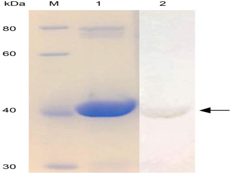
Fig. 3.
Purification of Streptococcus iniae alanine racemase. The enzyme was purified using Nickel ion affinity chromatography, analyzed by SDS-PAGE and western blotting. M: molecular weight standards; 1; 40 kDa SiAlr. 2: Western blotting analysis of the purified protein.
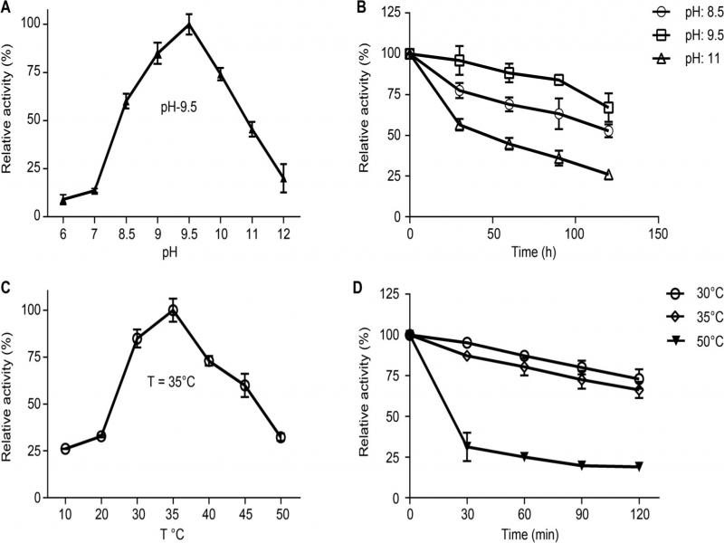
Fig. 4.
Effect of pH and temperature on the activity of SiAlr. (A) Optimal pH, (B) pH stability, (C) Optimal temperature, (D) Thermal stability.
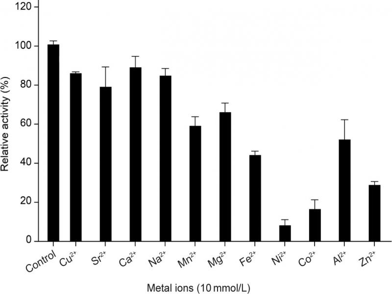
Fig. 5.
Effect of metals on SiAlr activity. The metal ions were at a concentration of 10 mM/L. The data were presented as mean ± SD from 3 independent determinations.
Table II
Effect of Hydroxylamine, DTT and PLP on SiAlr Activity.
| Chemical | Concentration (mM) | Relative activity (%) |
|---|---|---|
| None | 100 (0.7) | |
| Hydroxylamine | 0.1 | 21 (1.2) |
| 1 | 11 (0.8) | |
| 10 | 9 (1.4) | |
| DTT | 1 | 27 (3.1) |
| 3 | 2 (0.8) | |
| PLP | 0.01 | 56 (2.4) |
| 0.04 | 83 (1.5) | |
| 0.06 | 96 (2.7) |
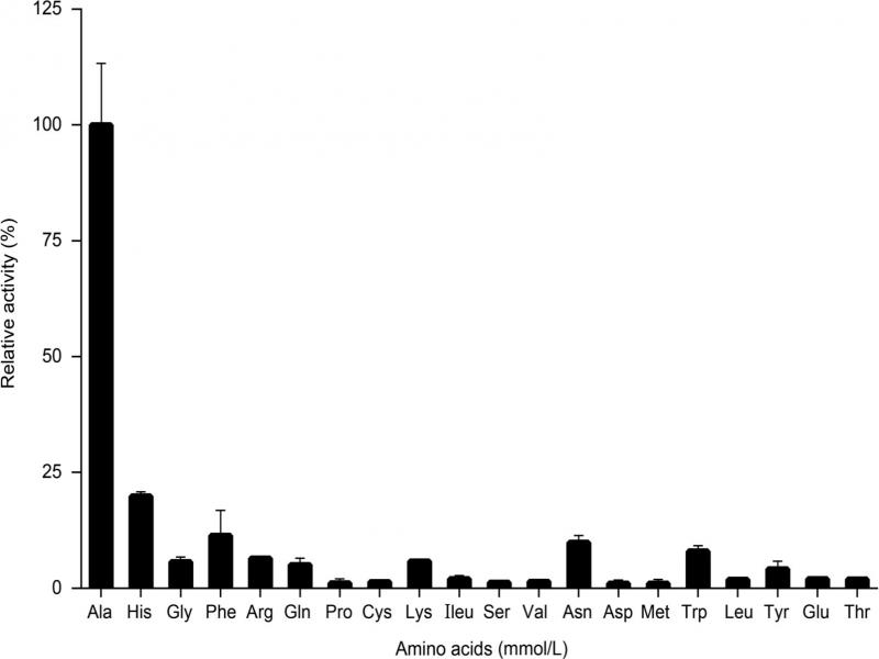
Fig. 6.
The substrate specificity of SiAlr. The relative activity of SiAlr for various L-Amino acids was determined at optimum pH and temperature. The data were presented as mean ± SD from 3 independent enzyme assays.
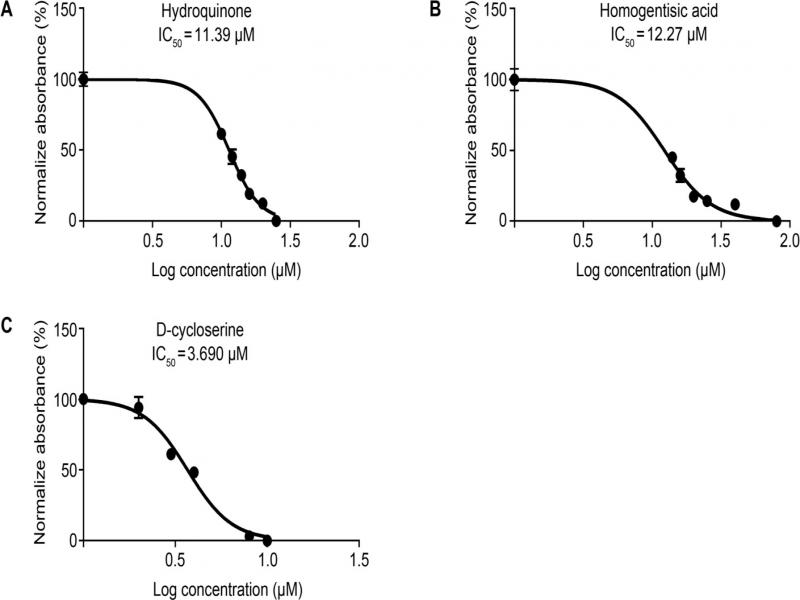
Fig. 7.
IC50 of the Streptococcus iniae alanine racemase inhibitors.
A: IC50 of Hydroquinone was 11.39 μM; B: IC50 of homogentisic acid was 12.27 μM; C: IC50 of D-cycloserine was 3.69 μM. The data shown are the means from three independent experiments.
Table III
The results of antimicrobial activity of homogentisic acid and hydroquinone inhibitors against numerous isolates of Gram-positive and Gram-negative bacteria.
| Organism | aMIC (μg/ml) | |
|---|---|---|
| Hydro-quinone | Homogentisic Acid | |
| Streptococcus iniae HNM-1 | 25 (2.3) | 200 (5.6) |
| Escherichia coli DH5α | 130 (7.9) | 210 (8.4) |
| Salmonella typhimurium | 150 (8.7) | 180 (11.4) |
| Staphylococcus aureus | 210 (13.7) | 250 (14.1) |
| Acinetobacter baumannii | 180 (11.5) | 210 (12.3) |
| Pseudomonas aeruginosa | 0 | 0 |