
1. Introduction
L. gibbosus (Linnaeus, 1758) is a freshwater and brackish-water species inhabiting the benthopelagic zone (Riede, 2004) at depths from the surface to 41 m (Scott & Crossman, 1973). Native to North America, the species is distributed from New Brunswick in Canada south to the Savannah River in Georgia, USA, and occurs in the Great Lakes, Hudson Bay (Red River), and upper Mississippi River basins, with introduced populations established in Pacific drainages from British Columbia to California. In Turkey, L. gibbosus was first recorded in the early 1980s and has since expanded widely across both European and Asian parts of the country (Ağdamar et al., 2015). Turkish populations exhibit unique haplotypes, low genetic diversity, and limited parasite communities, suggesting rapid adaptation and a potential competitive advantage through enemy release (Kvach et al., 2024). Recent records from Bayramiç Reservoir (December 2024) and Gökpınar Dam Lake (GDL) confirm ongoing range expansion (Kaya et al., 2025). Although trophic overlap with native species has been observed, studies suggest minimal immediate impacts on native fish condition, though continued monitoring is essential given the species’ demonstrated invasive potential (Karakuş et al., 2023). Individuals reach a maximum total length (TL) of 400 mm (Page & Burr, 2011) and a common length of 99 mm (Hugg, 1996), with a maximum recorded weight of 630 g (International Game & Fish Association, 1991) and a maximum reported age of 12 years (Hubbell, 1966). The species inhabits vegetated lakes, ponds, and quiet pools of creeks and small rivers (Page & Burr, 2011), feeding on small fishes, other vertebrates (Scott & Crossman, 1973), and fish eggs (Berg, 1965). Adults rarely form schools but occur in pairs or loose aggregations, while young individuals aggregate in larger schools (Jordan et al., 2009). In Europe, the species avoids swift waters and tolerates salinities up to 18.2 ppt (Kottelat & Freyhof, 2007), preying on a wide variety of invertebrates (Kottelat & Freyhof, 2007). Males construct nests in very shallow waters near the shore; the pair swims in a circular path over the nest, releasing eggs and sperm intermittently. Males guard the eggs for approximately 7 days (Gross & Sargent, 1985) and the young for about 11 days after hatching, then prepare the nest for another spawning with the same or different females (Scott & Crossman, 1973). In European waters, each male may spawn with several females in one nest and guard it until the larvae abandon it (Gross & Sargent, 1985). Fecundity reaches up to 1000 eggs per spawning event (Riehl & Baensch, 1991).
The study of morpho-anatomical abnormalities in fish dates back to the 16th and 17th centuries (Gudger, 1936), with early records documenting skeletal, nonskeletal, and pigmentation defects (Dawson, 1964, 1971). In recent decades, reports of skeletal deformities in wild fish populations have increased (e.g., Jawad et al., 2017a, on common pandora Pagellus erythrinus collected from the northern Aegean Sea, Turkey; Alpaslan et al., 2026, on Alburnus escherichii from the Sakarya River Basin, Türkiye; and Şavran et al., 2026, on Pseudorasbora parva, Pseudophoxinus battalgilae, and Oxynoemacheilus eregliensis sampled from the Gezende and Ermenek reservoirs in Türkiye), driven by anthropogenic stressors such as pollution and eutrophication (Leone et al., 2021), as well as heightened scientific interest in aquaculture-related bone malformations (Boglione et al., 2013a, 2013b; Ytteborg et al., 2012a, 2012b). These abnormalities, ranging from severe (fitness-impairing) to mild (minimal survival impact), can significantly affect fish morphology, growth, and survival (Gavaia et al., 2009; Jawad et al., 2016). Their occurrence in wild populations is typically rare, likely due to low prevalence or reduced viability under natural selection.
Monitoring skeletal deformities provides critical insights into environmental stress and aquaculture rearing conditions (Yershov, 2008). In Turkish freshwater systems, research on skeletal anomalies remains limited. Documented cases include ventral fin deformities in Capoeta damascina (Dağlı, 2008), kyphosis in Barbus pergamonensis (İnnal et al., 2019), and vertebral deformities in Barbus xanthos (Jawad & Güçlü, 2022), often linked to pollution. Similarly, L. gibbosus exhibits skeletal abnormalities in both natural and laboratory settings, frequently associated with environmental contaminants such as selenium (Lemly, 2014; Schmitt et al., 2019). High deformity rates in centrarchids, sometimes exceeding 50% in selenium-polluted waters (Gillespie & Baumann, 1986; Woock et al., 1987), underscore the ecological risks posed by toxicants.
Exposure to environmental pollutants induces morphological abnormalities and physiological stress in Lepomis sunfish. In the contaminated Flix reservoir (Spain), L. gibbosus exhibited significantly higher DELT anomalies (deformities, eroded fins, lesions, tumors) compared to reference sites (Benejam Vidal, 2008). Similarly, a Turkish study documented gill alterations, liver damage, muscle necrosis, and increased micronucleus formation (indicating genotoxic stress) in L. gibbosus from Çine Stream (Koca et al., 2005). Studies on the related longer sunfish (Lepomis megalotis) from metal-contaminated streams showed that lead, cadmium, and zinc exposure reduced vertebral calcium, phosphorus, and collagen (Lohner et al., 2001). Acute exposure of L. gibbosus to pesticides and pharmaceuticals has also been shown to cause gill alterations (Başimoğlu Koca & Kara, 2022). Collectively, these findings demonstrate that pollutants can lead to both external and internal deformities and histopathological changes in Lepomis sunfish.
Diagnostic advancements in detecting skeletal deformities have evolved significantly, with radiographic imaging emerging as a key tool. While mammography was originally designed for human breast imaging (O’Connell et al., 2018), its high-resolution X-ray capabilities have proven effective in detecting fine skeletal anomalies in fish (Güçlü et al., 2025; Jawad & Güçlü, 2022). Digital X-ray systems, including mammography units, offer superior spatial resolution for identifying ectopic mineralization, vertebral fusions, and spinal malformations (O’Connell et al., 2018). In aquaculture, techniques including dual-energy X-ray absorptiometry (DXA) have been validated for assessing skeletal defects in species such as Salmo salar (Drábiková et al., 2026; Fjelldal et al., 2012), though protocol adaptations are needed for fish-specific applications (Gjerde et al., 2005).
This study employs mammography-based radiography to document, for the first time, skeletal abnormalities in L. gibbosus from Turkish freshwater ecosystems. The findings reveal major deformities (vertebral fusion, spinal curvatures, caudal fin anomalies) and minor irregularities (malformed ribs, hemal spine deviations). By leveraging this diagnostic approach, the research contributes to (1) ecological health assessments, linking deformities to potential environmental stressors (e.g., pollution, habitat degradation); (2) aquaculture monitoring, validating noninvasive imaging techniques for deformity screening in farmed fish; and (3) methodological innovation, demonstrating the adaptability of medical radiography for ichthyological studies.
As skeletal malformations serve as biomarkers of environmental and husbandry-related stress, refining diagnostic techniques such as radiography is essential for advancing fish health research and conservation efforts.
2. Materials and methods
2.1. Description of the area
GDL, located in Denizli, holds the unique distinction of being Turkey’s first urban reservoir lake (Dikbaş, 2002) (Fig. 1). Formed geologically at the intersection of two active grabens—the NW– SE extending Gediz Graben and the E–W trending Menderes Graben—its structure was shaped by post-Neogene tectonic activity. The lake’s basin primarily consists of siltstone, sandstone, and marl units (Beyaz et al., 2007). Also referred to as the Vali Recep Yazıcıoğlu Reservoir, GDL lies within Denizli province in the Büyük Menderes River (BMR) basin. Initially constructed for irrigation, it became operational in 2004. The lake is fed by multiple sources, including Tekke and İğdeli streams, drainage channels, and surface runoff. With water levels fluctuating between 317 m and 336.2 m, GDL spans 1.98 km2 and boasts a storage capacity of 28.2 hm3 (U’hver et al., 1989). While the reservoir primarily supplies irrigation water, excess flow is discharged into the BMR via Çürüksu Stream. Two monitoring stations have been established within GDL: Station 1 (37°46′07″ N; 29°07′29″ E), located on the southwestern shore near the water inlet, features a gently sloping shoreline with moderate vegetation and a partially rocky, firm bottom. Station 2 (37°47′03″ N; 29°07′58″ E), positioned near the dam on the northern side, shares a similar substrate but has a steeper slope. The BMR, the largest river system in Western Anatolia, receives contributions from GDL and its tributary, Tekke Creek, within Denizli province (Yıldırım & Çetinkaya, 2024). Unlike stratified lakes, GDL maintains a single-layer current system, ensuring uniform oxygen distribution from surface to bottom, regardless of temperature or wind conditions (Dikbaş, 2002). This homogeneity was expected to support dense bottom vegetation and high organic matter accumulation. However, sediment sampling (2019–2020) revealed that only Station 1 exhibited a silt-mud substrate with sparse vegetation, while all other shorelines and bottom areas were completely barren.
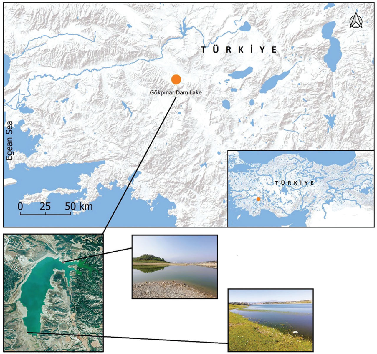
Figure 1
Map of GDL showing the sampling stations where L. gibbosus were collected. GDL, Gökpınar Dam Lake.
2.2. Fish samples
All experimental procedures involving animals were conducted in strict compliance with the animal welfare regulations, ethical guidelines, and policies mandated by the Republic of Türkiye. In May 2019, fifty individuals of L. gibbosus (Fig. 2) were collected using a combination of an experimental seine net and a multi-mesh gillnet. Among these, 16 exhibited varying degrees of skeletal deformities. Following anesthesia administration, detailed morphological measurements were taken with a dial caliper, accurate to the nearest millimeter. Each measurement followed a point-to-point protocol, excluding projections, in accordance with the standardized methods of Hubbs and Lagler (1958). TL was recorded as the linear distance from the snout tip to the posterior margin of the caudal fin, where the upper and lower lobes meet. Standard length (SL) was measured from the snout tip to the terminal end of the hypural complex. To assess skeletal anomalies, specimens underwent high-resolution radiographic imaging under optimized parameters (60–80 kV, 30–45 mAs, 2.1 s exposure time) using a Siemens Mammomat Inspiration (2013) mammography unit at Meddem Hospital’s Radiology Department in Isparta, Türkiye. Upon completion of the study, specimens were systematically catalogued (Catalogue no. UGYC-LGI-GBP-2019-20-A) and placed in the curated fish collection of the Faculty of Eğirdir Fisheries, Isparta University of Applied Sciences. Voucher specimens are accessible upon reasonable request to the first author.
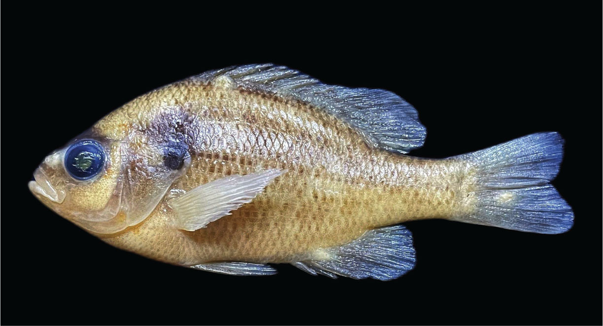
Figure 2
Normal specimens of L. gibbosus (73.6 mm SL). SL, Standard length.
2.3. Types of abnormalities
Skeletal anomalies were classified into two categories based on established criteria in fish skeletal biology (Davie et al., 2019; Witten et al., 2005; Ytteborg et al., 2012a, 2012b). Major abnormalities were defined as deformities affecting the axial skeleton—specifically the vertebral column and associated supporting structures—that compromise structural integrity, swimming performance, or long-term survival. These included vertebral coalescence, hypural bone deformation, and abdominal rib deformation. Minor deformities were defined as anomalies affecting peripheral or appendicular structures (fins and their supporting elements) that do not significantly impair locomotion or viability and may be subject to remodeling or compensation during growth. These included caudal fin ray deformity, hemal spine deformity, and parhypural bone deformity.
3. Results
3.1. Vertebral column regionalization and elements in examined fish
The vertebral column of teleost fishes was examined for regionalization based on the morphology of vertebral centra and their associated elements, including neural arches, hemal arches, parapophyses, and ribs (De Clercq et al., 2017). Following the refined regionalization scheme established for salmonids and applicable to other teleosts, the vertebral column was subdivided into distinct anatomical regions using radiographic hallmarks (Sankar et al., 2024). These regions were identified as postcranial (vertebrae lacking ribs), abdominal (vertebrae bearing ribs that articulate with parapophyses), transitional (vertebrae showing gradual phenotypic change from ribs to vestigial ribs and the presence of hemal arches), caudal (vertebrae with neural and hemal arches and spines), and ural (vertebrae with modified elements supporting the caudal fin) (De Clercq et al., 2017). Regional boundaries were determined based on the presence or absence of specific skeletal features visible on radiographs, including the modified parapophysis of the first transitional vertebra, the prominent hemal spine of the first caudal vertebra, and the separated hemal spine of the most cranial preural vertebra (Sankar et al., 2024).
To ensure a standardized and comparative framework for the skeletal anomalies observed in L. gibbosus, the deformities of the vertebral centra and their associated elements can be systematically categorized according to established morphological criteria. Following the detailed regionalization scheme for the teleost vertebral column (Arratia et al., 2001; De Clercq et al., 2017), the present study documented anomalies in both the vertebral centra (the main bony bodies) and their associated neural and hemal arches, spines, and ribs. The major deformity of the vertebral centra, vertebral coalescence, was confined to the caudal region, specifically involving the third and fourth preural vertebrae. This fusion is characterized by the loss of adjacent centra halves and their consolidation into a single, irregular bony mass with duplicated neural and hemal spines, a phenotype consistent with a localized remodeling process that has been previously described in salmonids (De Clercq et al., 2017; Witten et al., 2006). This pattern of localized fusion, affecting a few adjacent vertebrae, aligns with the ‘containment’ or ‘self-limiting’ model of vertebral fusion (Davie et al., 2019).
Deformities of the vertebral centra-associated elements were more diverse and included malformations of the hypural bones, abdominal ribs, hemal spines, and parhypural bone. The hypural bone deformation, characterized by an upward tilting of the entire caudal complex including the hypurals and uroneurals, represents a disruption in the late-stage development and ossification of these caudal-fin support structures. This integrated malformation suggests a potential error in the segmentation or ossification of the posterior vertebrae and their associated hypural elements (Arratia & Schultze, 1992; Arratia et al., 2001). The deformation of the abdominal ribs, where distal ends are misdirected anteriorly or posteriorly, indicates a developmental aberration in the patterning of these dermal bones, which, unlike vertebral centra, do not form through chondral ossification (De Clercq et al., 2017). Among the minor anomalies, the deformities of the hemal spines (curvature, notches) and the parhypural bone (right-angled curvature, displacement) are consistent with defects in elements that undergo chondral ossification (Fernandez & Gisbert, 2010). By categorizing these anomalies into centra versus centra-associated elements, the current study provides a clear, comparative record of skeletal pathology that can be directly compared with findings in other teleost species (Britz & Johnson, 2005; Johnson & Britz, 2010), thereby facilitating a standardized count and future meta-analyses of vertebral deformities in fishes.
The mammography of a normal L. gibbosus specimen is shown in Fig. 3. Across the 50 specimens examined, six types of skeletal deformities were documented, comprising three major and three minor categories. Among the major anomalies, abnormal abdominal ribs were the most prevalent, observed in four specimens (8%), followed by vertebral coalescence and deformed hypural bones, each observed in two specimens (4%). Among the minor deformities, caudal fin ray deformity and hemal spine deformity were each observed in two specimens (4%), while parhypural bone deformity was the least frequent, recorded in a single specimen (2%). Overall, skeletal abnormalities were present in 22% of the examined population (11 out of 50 specimens).
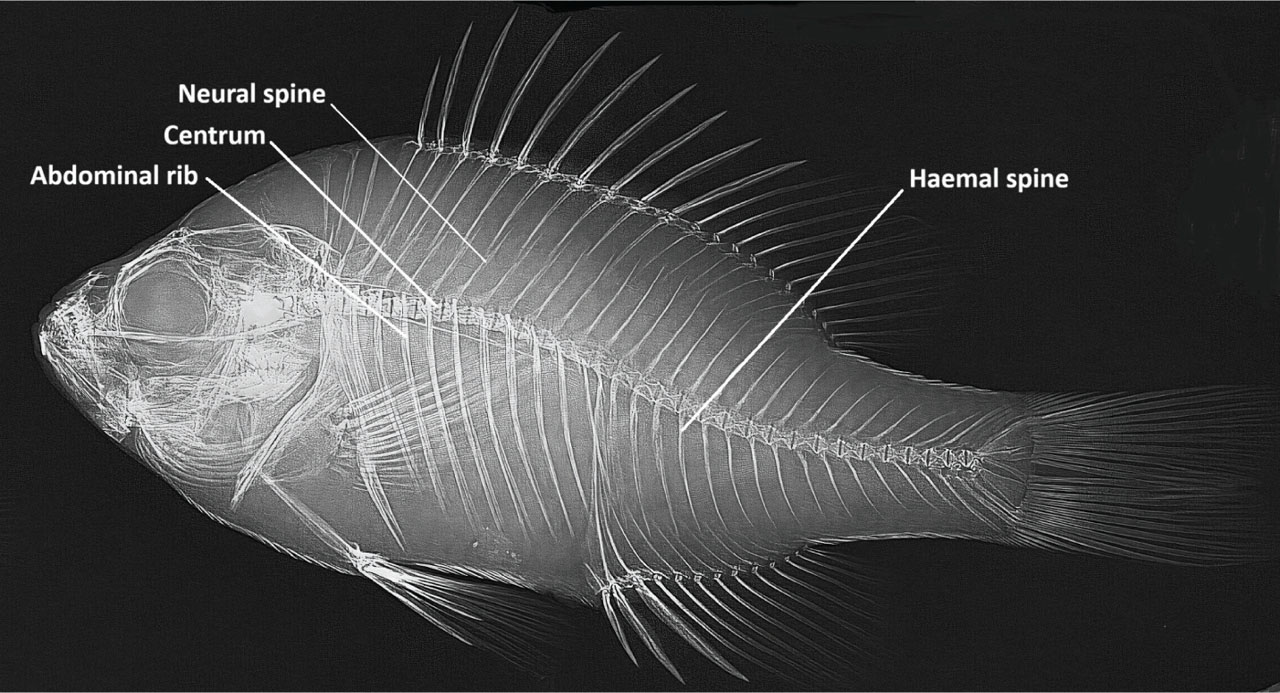
Figure 3
Mammography showing the normal skeletal system of L. gibbosus (36.3 mm SL). SL, Standard length.
3.2. Major abnormalities
3.2.1. Vertebral coalescence
Vertebral fusion is characterized by the abnormal union of two or more vertebrae. This condition typically involves the partial or complete loss or deformation of the vertebral centra, with the extent of the deformity varying from case to case.
A detailed examination of the two affected specimens of L. gibbosus revealed that the fusion specifically involved the third and fourth preural vertebrae (2/50, 4%). The anomaly presented as a loss of the posterior half of the fourth preural centrum and the anterior half of the third preural centrum. The remaining portions of these two vertebrae were fused into a single, irregularly sized bony element with two neural and hemal spines (Fig. 4).
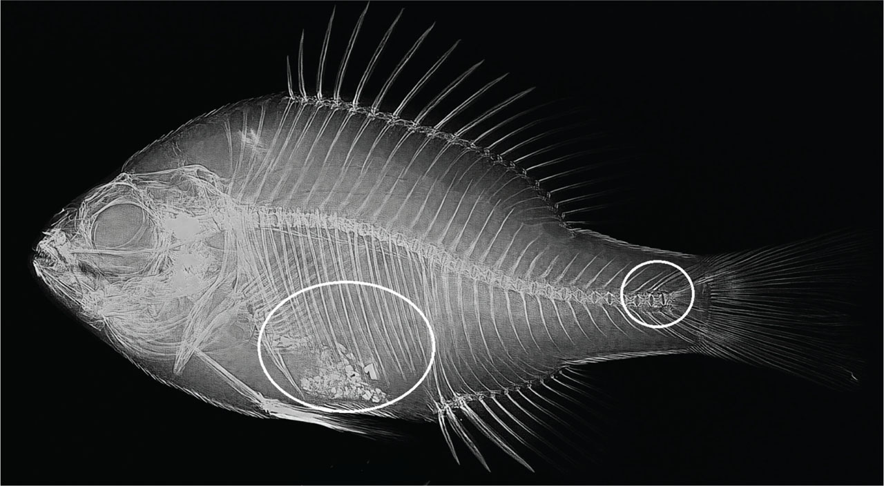
Figure 4
Specimen of L. gibbosus (36.3 mm SL) exhibiting a representative case of vertebral fusion in the caudal vertebrae and an abdominal rib deformity. SL, Standard length.
3.2.2. Deformation of the hypural bones
Two types of hypural bone deformity were observed in two specimens (2/50, 4%). In one specimen (76.6 mm SL, Fig. 5A), the following caudal fin elements appeared tilted upward compared to their normal position as shown in the radiograph of the normal specimen (Fig. 3): the first and second uroneural bones, the fourth and fifth hypural bones, all dorsal procurrent rays, all caudal fin rays of the upper lobe, and the two dorsal-most caudal fin rays of the lower lobe. In the second specimen (67.9 mm SL, Fig. 5B), a similar deformity was observed, with evident tilting of the caudal fin skeletal elements. However, in this specimen, the caudal fin rays themselves were not tilted and appeared normal compared to those of the normal specimen.
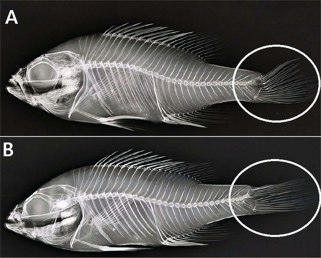
Figure 5
Deformities of the hypural bones in L. gibbosus. (A) Specimen at 76.6 mm SL; (B) specimen at 67.9 mm SL. SL, standard length.
3.2.3. Deformation of the abdominal ribs
This type of deformity was observed in four specimens (4/50, 8%). Misalignment was obvious along the entire length of all abdominal ribs. Instead of being directed ventrally, the distal ends of all abdominal ribs were directed either anteriorly or posteriorly (Figs. 4 and 6).
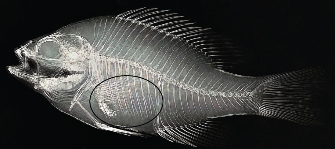
Figure 6
Abdominal rib deformity in a L. gibbosus specimen (75.4 mm SL), characterized by misalignment along the entire length of all ribs. SL, standard length.
3.3. Minor deformities
3.3.1. Caudal fin ray deformity
In this deformity, the caudal fin rays of both the upper and lower lobes were affected and observed in two specimens. In one specimen, the lower six caudal fin rays of the lower lobe were acutely curved downward. The fifth caudal fin ray (counting from the ventral side of the fins) showed a callus at its anterior one-third of its length (Fig. 7). In another specimen, all rays of the upper caudal fin were wavy in their posterior third, rather than straight as in the normal specimen. The two most ventral caudal fin rays were strongly waved in their posterior third. Additionally, the two uppermost caudal fin rays appeared wavy along their entire length (Fig. 8).
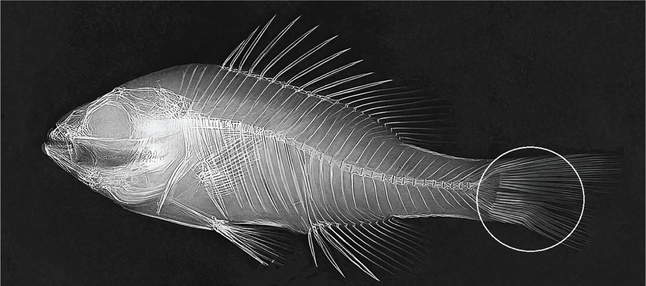
Figure 7
Caudal fin deformity in a L. gibbosus specimen (56.5 mm SL). The six ventral-most caudal fin rays are acutely curved downward. The fifth ray (counting ventrally) exhibits callus formation on the anterior third of its length. SL, standard length.
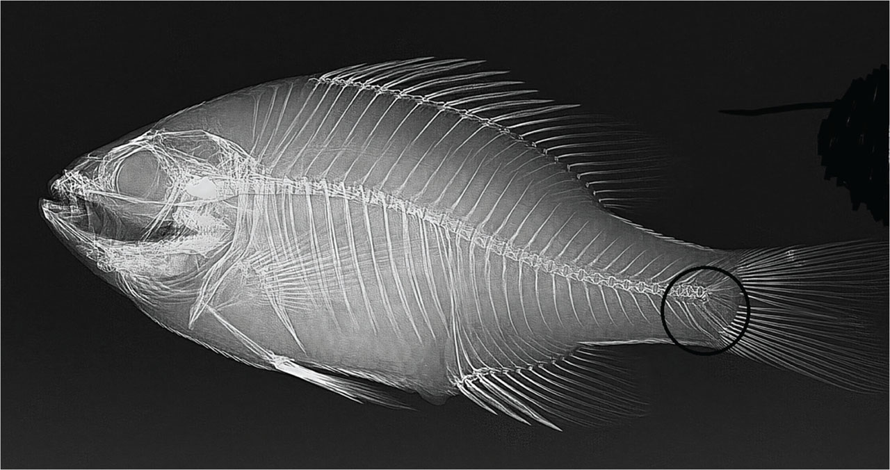
Figure 8
Hemal spine deformity in a L. gibbosus specimen (72.1 mm SL), where the hemal spine of the second preural vertebra had a posteriorly directed notch at its proximal end and was curved anteriorly at its distal end. SL, standard length.
3.3.2. Deformity of the hemal spine
This type of deformity was shown in two specimens; the hemal spines of the second, third, and fourth preural vertebrae were abnormal compared to those of the normal specimen. The hemal spine of the third preural vertebra was curved anteriorly at its mid part and posteriorly at its distal end (Fig. 8). In another specimen, the hemal spine of the second preural vertebra had a posteriorly directed notch at its proximal end and was curved anteriorly at its distal end (Fig. 9).
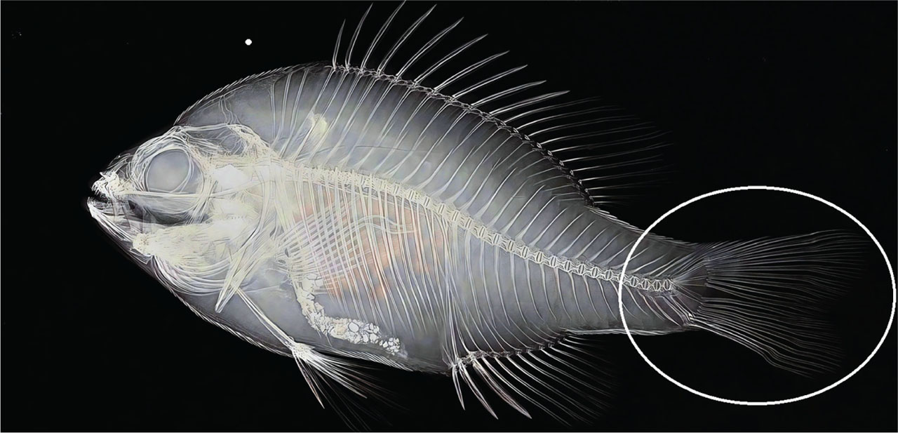
Figure 9
Skeletal deformities of the hemal spine, caudal fin rays, and parhypural bone in a L. gibbosus specimen (69.2 mm SL). SL, standard length.
3.3.3. Deformity of the parhypural bone
In this deformity, the parhypural bone was curved at its distal end, forming a right angle. In addition, it appeared displaced posteriorly, closer to the hemal spine of the second preural vertebra (Fig. 9).
4. Discussion
This investigation delivers the first documented evidence of spinal pathologies in a wild population of the teleost L. gibbosus from a freshwater habitat in Türkiye. The primary objective was the comprehensive diagnosis and characterization of skeletal deformities, thereby creating a reference benchmark for subsequent studies.
The present study documents a notable prevalence and diversity of skeletal deformities in a sample of L. gibbosus, revealing six distinct types of anomalies affecting the abdominal and caudal skeleton. The identification of three major abnormalities—vertebral coalescence, hypural bone deformation, and abnormal abdominal ribs—highlights significant disruptions in skeletal development. Vertebral fusion, particularly involving the preural vertebrae as observed here, is a well-documented abnormality often linked to disruptions during early ontogeny (Witten & Huysseune, 2009). Similarly, the misalignment of abdominal ribs and the tilting of the caudal complex suggest potential insults during the patterning and ossification processes of these elements. The co-occurrence of such severe malformations in a single population sample indicates the presence of underlying causative factors, whether hereditary or developmental, that merit further investigation.
The prevalence rates of skeletal deformities observed in the present study (22% overall) fall within the range reported for wild fish populations, though they are notably lower than those frequently documented in aquaculture settings, where rates can exceed 45% in certain production batches (Boglione et al., 2013a). The most prevalent major anomaly was the deformation of the abdominal ribs (8%), followed by vertebral coalescence and hypural bone deformation (each 4%). The higher prevalence of rib deformities compared to vertebral fusions is consistent with the findings of previous studies, which have suggested that ribs may be more sensitive to developmental perturbations than vertebral centra, particularly during early ossification (Jiménez-Guerrero et al., 2024). The localization of vertebral fusion to the caudal region (third and fourth preural vertebrae) is noteworthy, as the caudal vertebral column has been identified as a region of heightened susceptibility to developmental errors, potentially due to the complex morphogenetic processes involved in the formation of the caudal fin support structures (Haga et al., 2002; Witten et al., 2006). The minor deformities, including caudal fin ray deformity (4%), hemal spine deformity (4%), and parhypural bone deformity (2%), though less severe, are consistent with sub-lethal developmental disturbances that have been previously documented in teleosts (Boglione et al., 2013b; Koumoundouros, 2010). The overall prevalence of skeletal anomalies in this wild population of L. gibbosus provides a valuable baseline for future comparative studies and highlights the importance of radiographic screening for detecting internal deformities that may not be apparent through external examination alone (Gomes et al., 2017; Losada et al., 2014).
In addition to the major deformities, the presence of minor abnormalities affecting the caudal fin rays, hemal spines, and parhypural bone provides a more comprehensive picture of the skeletal instability within the studied group. While these minor defects may have a less pronounced impact on overall swimming performance compared to vertebral fusions, they are nonetheless critical biomarkers of developmental compromise. The waviness of fin rays and the aberrant curvature of spinal processes are consistent with sub-lethal developmental disturbances that can affect locomotor efficiency and long-term fitness (Boglione et al., 2013b). The spectrum of deformities recorded, from severe column malformations to finer anomalies in appendicular structures, underscores the value of L. gibbosus as a potential model for monitoring developmental health in wild fish populations.
Radiographic imaging, particularly mammography, is the gold-standard diagnostic technique for detecting skeletal deformities in fish, permitting a nondestructive and comprehensive analysis of the entire skeleton and its individual components (Fjelldal et al., 2007, 2009; Witten & Hall, 2003). This methodology consistently uncovers a critical diagnostic gap between external and internal examination. For instance, comparative studies reveal that a substantial proportion of visually ‘normal’ fish harbor concealed anomalies, with one study finding 72% undetected anomalies and 46% vertebral body abnormalities (Gomes et al., 2017). This pattern is corroborated by work on Senegalese sole, in which approximately 76% of morphologically normal individuals exhibited internal deformities (Losada et al., 2014), collectively demonstrating the low sensitivity of macroscopic assessment.
The long-term implications of subvisual deformities extend far beyond their initial lack of visibility. While such anomalies may not compromise immediate market appeal for commercial fish species (Deschamps et al., 2008), they are often progressive in nature. Studies have documented that these skeletal irregularities can advance in later developmental stages (Fjelldal et al., 2007; Witten et al., 2006), potentially escalating into severe, externally manifested conditions that detrimentally affect the fish at later developmental stages (Deschamps et al., 2008).
Vertebral fusion, a severe skeletal malformation, is pathologically defined by the abnormal union of two or more vertebrae (Lauder, 1982). This process fundamentally disrupts normal skeletogenesis, typically manifesting as the partial or complete loss, resorption, or severe deformation of the intervertebral connective tissues and the vertebral centra themselves (Witten & Huysseune, 2009). The resulting fused structure often forms a single, irregularly shaped bony mass, which can incorporate duplicated or misshapen neural and hemal arches (Boglione et al., 2013b). This pathological consolidation compromises the flexibility and mechanical integrity of the skeleton, potentially leading to impaired locomotion, reduced fitness, and secondary health complications (Sfakianakis et al., 2004).
The developmental trajectory of spinal deformities, such as vertebral fusion, has been systematically classified in prior research. Notably, the four-stage model of vertebral fusion proposed by Ytteborg et al. (2012a, 2012b) provides a particularly relevant framework for the anomalies documented in the present study. This model describes a progressive pathogenic sequence that commences with the invasion of disorganized, proliferating cells into the growth zones of adjacent vertebral bodies. This initial cellular disruption represents a critical first step, ultimately resulting in the failure of normal segmentation and the subsequent formation of a fused bony mass. The fundamental mechanism underpinning this pathology—a disruption in the balance between programmed cell death (apoptosis) and uncontrolled cellular proliferation—represents a conserved pathogenic pathway well-documented in mammalian skeletal malformations (Tozer & Duprez, 2005). This observation suggests that, despite the vast evolutionary distance separating teleosts and mammals, the core regulatory processes governing vertebral development and dysmorphogenesis are shared across vertebrate lineages (Hall & Miyake, 1995).
The findings of the present investigation suggest that the observed vertebral fusion in L. gibbosus conforms to a pathogenic model of localized dysregulation. In this scenario, the affected vertebral centra do not merely join but undergo a comprehensive remodeling process, ultimately consolidating into a single, well-defined, and morphologically stable bony element. This specific type of fusion was first characterized in S. salar, where it was distinguished from more disruptive malformations (Witten et al., 2006). Our results thus provide compelling cross-species validation for this developmental model. The biological principle of this fusion type appears to be a conserved vertebrate phenomenon, as it is also documented in human medicine, where it can alter spinal biomechanics and lead to secondary compensatory deformities in adjacent segments (Leivseth et al., 2005). Critically, this study identifies the first documented case of this fusion scenario within the species L. gibbosus, expanding the known taxonomic range of this specific pathological pathway.
The localized model of vertebral fusion finds further compelling support in a study of farmed Oncorhynchus tshawytscha conducted by Davie et al. (2019) in New Zealand. Their research documented a consistently low average number of vertebrae implicated in each fusion event, a morphological pattern they directly attributed to a high prevalence of the localized fusion type. A key conclusion from their work was that, irrespective of the chronicity or duration of the condition, the fusion process is typically self-limiting, involving only a few adjacent vertebrae and failing to propagate progressively along the spinal column to incorporate additional elements. This established principle aligns precisely with the observations in the present study, wherein all recorded fusion anomalies in the examined specimens were strictly confined to a pair of adjacent vertebrae. This remarkable consistency across geographically and phylogenetically distinct teleost species not only reinforces the validity of this model but also suggests it may represent a fundamental and conserved pathway in piscine skeletal repair or dysmorphogenesis (Witten & Huysseune, 2009).
The pathogenesis of vertebral fusion is increasingly understood as a disruption of fundamental developmental pathways governing skeletal patterning. A pivotal study by Haga et al. (2009) established a direct link between these malformations and primary defects in the embryonic processes of notochord segmentation and subsequent vertebral centrum differentiation. These meticulously orchestrated events are highly vulnerable to perturbation; for instance, accelerated skeletogenesis, such as that triggered by exogenous exposure to vitamin A, can desynchronize development and precipitate such errors (Mazurais et al., 2008). Beyond early development, the integrity of the spine is maintained by notochord-derived tissues.
Experimental research utilizing vitamin A-induced teratogenesis has been instrumental in elucidating the mechanisms behind vertebral fusion. Seminal work by Haga et al. (2011) demonstrated that hypervitaminosis A produces distinctively malformed vertebrae, characterized by fused centra of increased length and the anomalous presence of duplicated neural and hemal arches. Critically, this teratogenic effect was established as dose-dependent in Japanese flounder, with severity escalating alongside concentration (Haga et al., 1999). The primary causative agent is attributed to retinoic acid (RA), a potent bioactive metabolite of vitamin A. High concentrations of RA are known to accumulate in live feeds such as rotifers and Artemia nauplii, which are commonly used in aquaculture (Takeuchi et al., 1998). A key finding from this line of research is the regional susceptibility of the vertebral column; the caudal region exhibits a particular vulnerability to the disruptive effects of vitamin A and RA on skeletogenesis (Haga et al., 2002). This established site-specific sensitivity is directly consistent with the present findings in L. gibbosus, where the observed fusion was localized to the caudal vertebrae.
Deformities of the hypural bones, which form the structural foundation for the attachment of the caudal fin rays, represent a significant skeletal pathology in teleost fish, often leading to impaired swimming performance and reduced survival. In general, these anomalies can manifest as malformations, fusions, or improper angling (tilting) of the hypural plates, which disrupts the precise biomechanics of the caudal fin (Boglione et al., 2013b). In the specific case of L. gibbosus, the deformity presents as a distinct upward tilting of the entire caudal complex, involving not only the hypural bones but also associated elements such as the uroneurals, resulting in a misalignment of the fin rays. This type of integrated deformity suggests a disruption during the late stages of caudal skeletogenesis, potentially originating from errors in the segmentation or ossification of the posterior vertebrae and the hypural elements that develop in close association with them (Witten et al., 2006). The consistent presentation of this tilted morphology in L. gibbosus indicates a potential species-specific susceptibility or a common, unidentified teratogenic insult affecting the development of this critical locomotor structure.
The etiology of hypural bone deformities in teleost fish is recognized as multifactorial, stemming from an interplay of genetic predispositions and developmental disruptions. In the present study, temperature fluctuations during critical windows of larval development, sublethal hypoxia, and exposure to teratogenic chemicals are significant contributors (Sfakianakis et al., 2004). A prominent example is the disruptive role of elevated levels of RA, a vitamin A metabolite, which can directly interfere with notochord segmentation and the subsequent formation of the hypural plates, leading to fusions and malalignments (Haga et al., 2011). Often, these factors are not mutually exclusive, as developmental stressors can exacerbate underlying genetic weaknesses, culminating in the expression of severe caudal deformities.
Abdominal rib deformities were documented in four specimens of L. gibbosus, with the affected individuals exhibiting a misalignment along the entire length of all abdominal ribs. Instead of being directed ventrally, the distal ends of all abdominal ribs were directed either anteriorly or posteriorly. The foundational anatomy of teleost epipleural ribs, intermuscular bones, and associated ligaments was comprehensively detailed by Patterson and Johnson (1995), with a comparable deformity subsequently documented in Oncorhynchus mykiss by Gislason et al. (2010). Critically, however, the rib deformities identified in the present study represent the first documented instances within L. gibbosus. This finding is particularly noteworthy in light of the recent compilation by Lyall et al. (2024), which catalogued cyprinid species with rib anomalies yet did not include L. gibbosus. Further contextualizing rib pathologies, Lyall et al. (2024) described distinct curled deformations in the pleural ribs of Salmo trutta and associated vertebral anomalies, positing that the underlying etiological mechanisms may have broad implications for skeletal integrity. In a complementary study, Jiménez-Guerrero et al. (2024) identified generalized radiolucency and axis deviation as the two predominant rib abnormalities in salmon, noting that ribs demonstrated greater sensitivity to dietary shifts during smoltification than vertebral elements. From a pathophysiological perspective, generalized radiolucency—often indicative of aberrant development, degeneration, or osteomalacia—can confer abnormal flexibility to the ribs (Khurana, 2009). This paradoxical state may reduce the likelihood of brittle fracture while simultaneously predisposing the bone to plastic deformation. It is crucial to acknowledge, however, that this relationship is not universal, as other pathologies such as osteomyelitis and osteoporosis can significantly increase skeletal fragility and fracture risk (Jiménez-Guerrero et al., 2024).
A singular specimen of L. gibbosus exhibited a distinctive caudal fin ray deformity, affecting both the upper and lower lobes. The anomaly was characterized by a pronounced waviness, contrasting with the straight rays of a normal specimen. Specifically, all rays in the upper lobe displayed this undulating morphology in their posterior third. The two most ventral rays were strongly waved in this region, while the two uppermost rays were slightly wavy in their anterior third. The remaining three rays exhibited this waviness along their entire length. Caudal fin deformities, documented in both wild and cultured populations, manifest with varying severity and phenotypes. These range from fin structure and ray agenesis to lateral bending of the entire complex or even caudal fin duplication (Fragkoulis et al., 2020; Koumoundouros, 2010). The literature suggests causative factors are diverse, often implicating rearing conditions for aquaculture species and nutritional deficiencies for wild fish (Boglinoe et al., 2012; Mazurais et al., 2009). While a genetic basis has been proposed (Fragkoulis et al., 2020), research in this area remains limited, with foundational work including studies on O. niloticus (Mair, 1992) and Cyprinus carpio (Kocour et al., 2006). Such deformities often originate from an abnormal bending of the notochord’s posterior tip during the yolk-sac stage, a critical period preceding caudal skeleton development (Koumoundouros et al., 1997). Consequently, the affected specimen in this study could have sustained this malformation for years. While nonfatal and not necessarily impeding essential biological functions such as feeding (Ribeiro-Prado et al., 2008), it is plausible that the deformity subtly compromised locomotor efficiency. This is supported by observations that, while the pectoral fins were morphologically pristine, the caudal fin lacked its characteristic symmetrical shape, a key feature for propulsion (Abed et al., 2024).
The remaining two minor skeletal deformities observed are the deformity in the hemal spine of the caudal vertebrae, observed in only one specimen, and the deformity of the parhypural, also observed in one specimen of L. gibbosus. In the deformity of the hemal spines, the hemal spines of the second, third, and fourth preural vertebrae were abnormal compared to those of the normal specimen. The hemal spine of the second preural vertebra was curved posteriorly at its distal end. The hemal spine of the third preural vertebra was curved anteriorly instead of being straight, while that of the fourth preural vertebra had a posteriorly directed notch at its proximal end and was curved anteriorly at its distal end. In the deformity of the parhypural bone, this bone appeared to be curved at its distal end, forming a right angle. In addition, it appeared displaced posteriorly, closer to the hemal spine of the second preural vertebra.
The etiology of these minor abnormalities is likely similar to those agents that induce skeletal deformities in various fish species, as reported by several authors (Abed et al., 2024). Fernandez and Gisbert (2010) demonstrated that skeletal structures undergoing chondral ossification, such as neural and hemal spines, epurals, parhypurals, and hypurals, are more sensitive to elevated dietary vitamin A levels than dermal bones, such as vertebral bodies. More recently, Fjelldal et al. (2018a, 2018b) observed fracture and fracture repair in neural and hemal spines in Gadus morhua. In this species, fractures healed normally through callus formation and subsequent remodeling. Some Labrus bergylta specimens exhibited abundant callus formation, larger than those reported by Fjelldal et al. (2018a, 2018b), but like those seen in Melanogrammus aeglefinus (Jawad et al., 2018) and Pomadasys stridens (Jawad et al., 2013).
In conclusion, this study establishes a critical baseline by providing the first comprehensive report of diverse skeletal deformities in a wild population of L. gibbosus. The spectrum of major and minor anomalies identified, particularly the vertebral fusions conforming to a conserved localized model, strongly points to underlying developmental disruptions. These findings position L. gibbosus as a potential model for studying skeletal development, and the detailed characterization provided here forms an essential reference for future investigations into the mechanisms underlying vertebral and skeletal deformities in freshwater fish populations.
Acknowledgments
We would like to express our gratitude to Dr. Salim Serkan Güçlü and Dr. Sera Övgü Kabadayi Yildirim from Isparta University of Applied Sciences for their assistance in the field and laboratory work for the first author. We also extend our thanks to Dr. Salim Serkan Güçlü for his help in performing mammography for the specimens.
Notes
[2] Contributed by Author contributions
Ufuk Gürkan Yıldırım: Observation, collecting fish specimens, performing mammography imaging, and reading the first draft of the manuscript.
Osman Çetinkaya: Observation, formal analysis, and methodology.
Laith A. Jawad: Conceptualization, formal analysis, investigation, methodology, project administration, supervision, validation, visualization, writing original draft, writing review, and editing.
[3] Conflicts of interest Conflict of interest
The authors declare that they have no known competing financial interests or personal relationships that could have appeared to influence the work reported in this article.