Figure 1
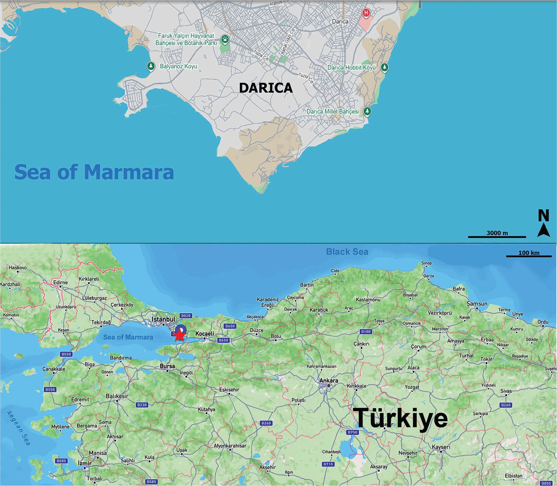
Figure 2
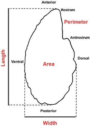
Figure 3
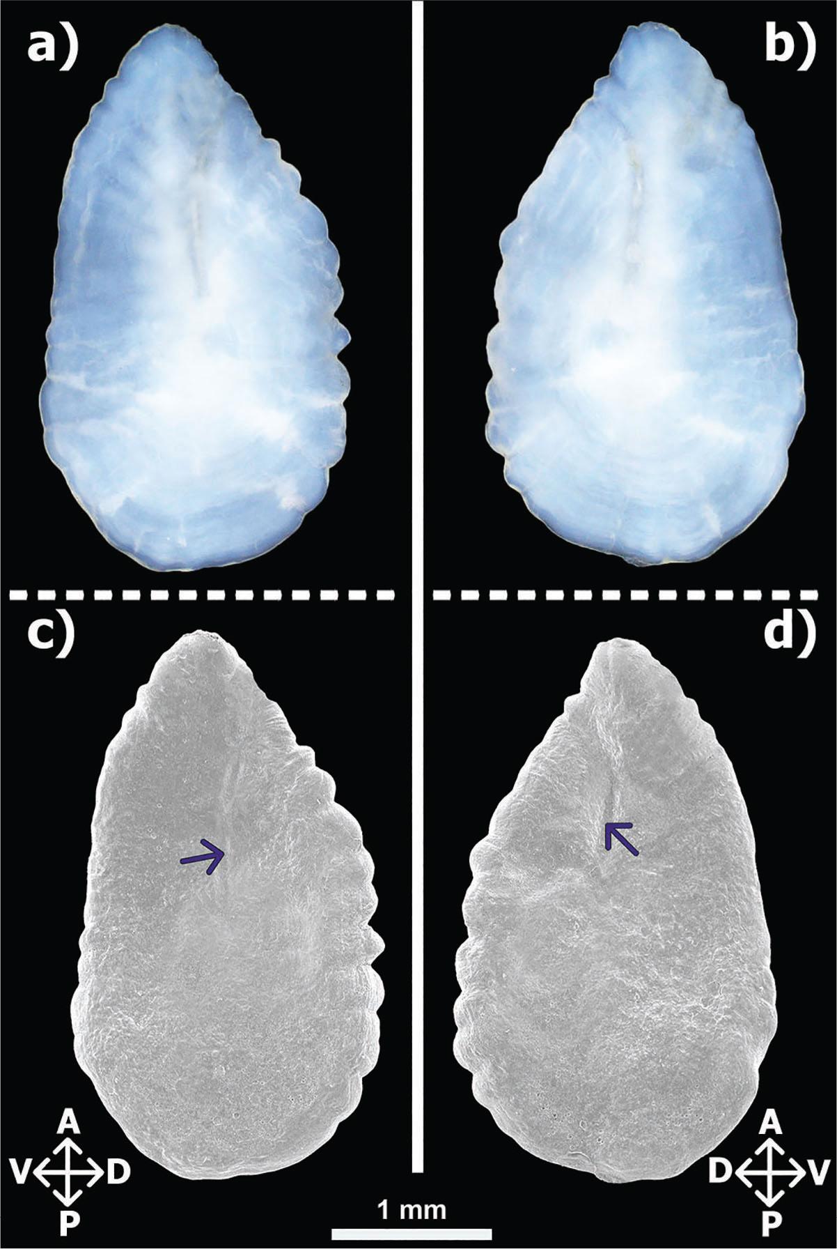
Figure 4

Figure 5
Figure 6

Figure 7

Basic measurements of Belone belone samples collected from the Darıca coast (Sea of Marmara, Türkiye) and comparisons of their abnormal and normal otolith morphometric data
| W (g) | TL (cm) | Sex | Side | Otolith Weight (g) | Otolith Area (mm2) | Otolith Perimeter (mm) | Otolith Length (mm) | Otolith Width (mm) | |
|---|---|---|---|---|---|---|---|---|---|
| Abnormal Samples | 19.2 | 25.9 | ♂ | Left | 0.0016 | 3.2019 | 8.0820 | 2.7729 | 1.6094 |
| Right | 0.0019 | 2.9783 | 7.2769 | 2.7036 | 1.5305 | ||||
| 69.4 | 39.0 | ♀ | Left | 0.0033 | 5.4039 | 11.1767 | 3.8532 | 2.1196 | |
| Right | 0.0037 | 5.2827 | 10.8174 | 3.9054 | 1.9942 | ||||
| 69.6 | 39.8 | ♀ | Left | 0.0050 | 6.3525 | 12.3690 | 4.1205 | 2.2615 | |
| Right | 0.0052 | 5.6110 | 11.3466 | 3.9115 | 2.1118 | ||||
| 115.8 | 44.2 | ♂ | Left | 0.0084 | 7.6115 | 12.4390 | 4.2390 | 2.4191 | |
| Right | 0.0079 | 7.6756 | 12.5602 | 4.3766 | 2.3989 |