
Figure 1.
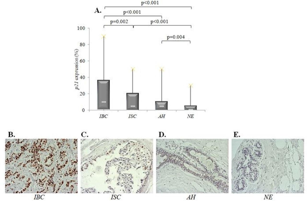
Figure 2.

Figure 3.

Figure 4.
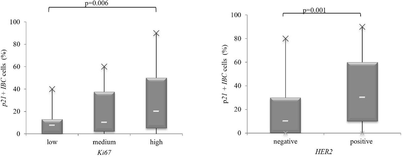
Figure 5.

Figure 6.

Figure 7.
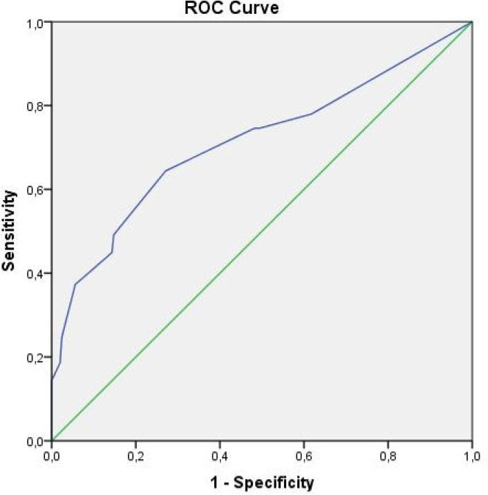
Figure 8.

Clinicopathological characteristics of breast cancer
| Variables | N | % | |
|---|---|---|---|
| Side | left | 66 | 44.9 |
| right | 81 | 55.1 | |
| Histological type | lobular | 18 | 12.4 |
| ductal | 123 | 84.8 | |
| other | 4 | 2.8 | |
| Histological grade | HG1 | 17 | 11.9 |
| HG2 | 73 | 51 | |
| HG3 | 53 | 37.1 | |
| Nuclear grade | NG1 | 17 | 15.2 |
| NG2 | 64 | 57.1 | |
| NG3 | 31 | 27.7 | |
| Mitotic index | grade 1 | 19 | 42.2 |
| grade 2 | 20 | 44.4 | |
| grade 3 | 6 | 13.3 | |
| Tumor necrosis | absent | 26 | 21.7 |
| present | 94 | 78.3 | |
| Desmoplasia | low | 17 | 16.3 |
| medium | 55 | 52.9 | |
| high | 32 | 30.8 | |
| Periductal elastosis | low | 19 | 20.0 |
| medium | 20 | 44.4 | |
| high | 16 | 35.6 | |
| Perineural invasion | absent | 101 | 68.7 |
| present | 46 | 31.3 | |
| Lymphatic invasion | absent | 72 | 48.9 |
| present | 75 | 51.1 | |
| Vascular invasion | absent | 113 | 76.9 |
| present | 34 | 23.1 | |
| HER2 | negative | 115 | 79.3 |
| positive | 30 | 20.7 | |
| Ki67 | low | 30 | 20..9 |
| medium | 42 | 29.4 | |
| high | 71 | 49.7 | |
| Molecular subtypes | Lum A | 30 | 20.4 |
| Lum B | 76 | 51.7 | |
| HER2 + | 19 | 12.9 | |
| TNBC | 22 | 15 | |
| T status | T1 | 48 | 35.8 |
| T2 | 64 | 47.8 | |
| T3 | 9 | 6.7 | |
| T4 | 13 | 9.7 | |
| N status | N0 | 50 | 37.3 |
| N1 | 48 | 35.8 | |
| N2 | 19 | 14.2 | |
| N3 | 17 | 12.7 | |
Association between p21 expression in IBC and examined clinicopathological characteristics_
| Variables | p21 cut off 7.5% | Chi-Square | p | ||
|---|---|---|---|---|---|
| − | + | ||||
| Mononuclear infiltrate | absent | 1 (9.1%) | 3 (5.8%) | 0.666 | 0.881 |
| low | 4 (36.4%) | 20 (38.5%) | |||
| medium | 5 (45.5%) | 20 (38.5%) | |||
| high | 1 (9.1%) | 9 (17.3%) | |||
| Histological type | lobular | 1 (6.7%) | 5 (6.7%) | 0.622 | 0.733 |
| ductal | 14 (93.3) | 67 (89.3) | |||
| other | 0 (0.0%) | 3 (4.0%) | |||
| Histological grade | HG1 | 2 (12.5%) | 8 (11.0%) | 0.364 | 0.834 |
| HG2 | 7 (43.8%) | 38 (52.1%) | |||
| HG3 | 7 (43.8%) | 27 (37.0) | |||
| Nuclear grade | NG1 | 0 (0.0%) | 6 (9.7%) | 2.096 | 0.351 |
| NG2 | 7 (53.8%) | 37 (59.7%) | |||
| NG3 | 6 (46.2%) | 19 (30.6%) | |||
| Tumor necrosis | absent | 2 (15.4%) | 17 (27.0%) | 0.278 | 0.598 |
| present | 11 (84.6%) | 46 (73.0%) | |||
| Perineural invasion | absent | 13 (81.3%) | 49 (64.5%) | 1.015 | 0.314 |
| present | 3 (18.8) | 27 (35.5%) | |||
| Lymphatic invasion | absent | 5 (31.3%) | 37 (48.7%) | 0.993 | 0.319 |
| present | 11 (68.8%) | 39 (51.3%) | |||
| Vascular invasion | absent | 12 (75.0%) | 57 (75.0%) | 0.000 | 1.000 |
| present | 4 (25.0%) | 19 (25.0%) | |||
| Molecular subtypes | Lum A | 1 (6.3%) | 10 (13.2%) | 11.490 | 0.009 |
| Lum B | 8 (50.0%) | 42 (55.3%) | |||
| HER2 + | 0 (0.0%) | 15 (19.7%) | |||
| TNBC | 7 (43.8%) | 9 (11.8%) | |||
| HER2 | negative | 16 (100.0%) | 49 (66.2%) | 5.895 | 0.015 |
| positive | 0 (0.0%) | 25 (33.8%) | |||
| Ki67 | low | 2 (13.3%) | 10 (13.5%) | 1.075 | 0.584 |
| medium | 6 (40.0%) | 20 (27.0%) | |||
| high | 7 (46.7%) | 44 (59.5) | |||
| T status | T1 | 3 (20.0%) | 22 (32.4%) | 2.924 | 0.404 |
| T2 | 9 (60.0%) | 34 (50.0%) | |||
| T3 | 2 (13.3%) | 3 (4.4%) | |||
| T4 | 1 (6.7%) | 9 (13.2%) | |||
| N status | N0 | 4 (26.7%) | 23 (33.3%) | 1.504 | 0.681 |
| N1 | 8 (53.3%) | 26 (37.7%) | |||
| N2 | 1 (6.7%) | 10 (14.5%) | |||
| N3 | 2 (13.3%) | 10 (14.5%) | |||