
1. INTRODUCTION
Mature peripheral T-cell lymphomas (PTCLs) are a heterogeneous group of lymphoid malignancies that generally follow an aggressive clinical course and are associated with poor outcomes. Globally, non-Hodgkin lymphoma accounts for approximately 544,000 new cases each year, with PTCLs representing approximately 10–15% of cases, although their frequency varies considerably across geographic regions [1,2]. Despite marked differences in their biology and clinical presentation, most PTCL subtypes continue to have poor long-term outcomes, with 5-year overall survival rates of approximately 30–40% in most series [3,4]. Overall, survival remains inferior to that achieved in aggressive B-cell lymphomas [5].
Progress in the treatment of PTCL has also been relatively slow. Although several new agents have entered clinical practice, conventional multiagent chemotherapy remains the basis of first-line treatment for many PTCL subtypes, and some treatment strategies are still adapted from those developed for aggressive B-cell lymphomas [6]. Better disease specific therapies are clearly needed, but so are biomarkers that can improve risk assessment and help identify patients with distinct disease biology.
Efforts to improve prognostic stratification have increasingly combined clinical features with biological markers. Adult T-cell leukemia/lymphoma (ATLL) is a good example. The ATLL Prognostic Index (ATL-PI) combines clinical variables, including Ann Arbor stage and performance status, with biological parameters such as soluble interleukin-2 receptor (sIL-2R) levels to stratify patients with acute and lymphoma type ATLL [7]. A prognostic model proposed in a North American cohort incorporated readily available parameters such as white blood cell and lymphocyte counts, corrected serum calcium, and lymphadenopathy [8]. In chronic and smoldering ATLL, sIL-2R has also been incorporated into prognostic assessment [9]. The inclusion of immune related parameters in these models raises the possibility that circulating cytokines and related biomarkers may provide additional information about disease behavior.
Compared with B-cell malignancies, progress in understanding the molecular biology of mature T-cell neoplasms has historically been slower, particularly in areas such as molecular characterization, intracellular signaling, and the interaction between malignant cells and their microenvironment [10,11]. At the same time, advances in cancer immunology have shown that the tumor microenvironment is not simply a background in which malignant cells grow, but a complex network of immune cells, stromal components, and soluble mediators that can either promote or restrain tumor development [12,13]. This interaction is particularly relevant in T-cell lymphomas because the malignant cells themselves arise from the immune system and can both respond to and modify immunoregulatory signals. Cytokines involved in T-cell activation and immune regulation may therefore offer insight into the biological state of these diseases.
IL-2 and IL-10 are immunoregulatory cytokines with established relevance to T-cell biology. IL-2 supports effector T-cell and natural killer cell activation while also being essential for regulatory T-cell homeostasis, and the IL-2/IL-2 receptor axis has prognostic relevance in ATLL [7,9,14,15]. IL-10 suppresses antigen presenting cell and pro-inflammatory functions and may contribute to the immunosuppressive microenvironment of T-cell lymphomas; elevated IL-10 has been associated with adverse outcomes in AITL, while experimental data support IL-10/STAT3/IRF4 signaling in ATLL [16,17,18,19,20,21,22]. In the present study, IL-2 and IL-10 were retained as descriptive contextual cytokines, whereas the survival analysis focused on IL-35.
Interleukin-35 (IL-35) is a heterodimeric immunosuppressive member of the IL-12 cytokine family, composed of the IL-12α (p35) and Epstein–Barr virus-induced gene 3 (EBI3) subunits [23,24]. It suppresses effector T-cell responses and promotes regulatory immune activity. Tregs are considered an important source of IL-35, although regulatory B cells (Bregs) and tolerogenic dendritic cells (tolDCs) can also produce it [24,25,26]. Within the tumor microenvironment, IL-35 has been linked to several processes that may facilitate tumor progression, including the expansion of regulatory immune cell populations, suppression of effector T-cell function, tumor-cell survival, and angiogenesis [27,28].
The role of regulatory immune mechanisms in T-cell lymphomas, however, appears to be more complex than in many solid tumors. In solid malignancies, increased intratumoral Treg infiltration is commonly associated with suppression of antitumor immunity. In lymphoma, the picture is less consistent, and studies have reported heterogeneous associations between Treg infiltration and outcome, including paradoxically favorable associations in some PTCL subtypes [29,30]. This is particularly relevant to IL-35 because of its close relationship with Treg biology. Rather than having a uniformly adverse effect, regulatory immune pathways may have different biological and prognostic implications depending on the specific T-cell lymphoma and its immune microenvironment [27,28,29,30].
These questions are also relevant in the Romanian clinical setting. Romania is a recognized European focus of human T-cell leukemia virus type 1 (HTLV-1) infection, and ATLL therefore represents a clinically relevant part of the spectrum of mature T-cell neoplasms encountered in this population [31,32]. Acute and lymphoma type ATLL have particularly aggressive clinical courses, while mature T-cell lymphomas as a group continue to have poorer outcomes than aggressive B-cell lymphomas [3,4,5]. Circulating immune biomarkers that reflect the interaction between malignant T cells and the host immune response could therefore complement established clinicobiological prognostic factors and potentially improve risk stratification.
In this exploratory study, we measured circulating IL-2, IL-10, and IL-35 at diagnosis in patients with mature T-cell neoplasms and examined their relationship with clinical outcome, with a primary focus on the association between plasma IL-35 and overall survival. We hypothesized that circulating immunoregulatory cytokines may reflect clinically relevant aspects of tumor–immune interaction in these heterogeneous malignancies.
2. MATERIALS AND METHODS
2.1. Study Cohort, Plasma Collection, and Cytokine Measurement
Diagnostic plasma samples included in the study were collected between 2020 and August 2025. This exploratory, non consecutive cohort comprised 20 adult patients with histologically confirmed mature T-cell neoplasms selected according to the availability of stored diagnostic EDTA plasma. Inclusion required: (i) a histologically confirmed mature T-cell neoplasm; (ii) an EDTA plasma sample obtained at diagnosis and available in sufficient quantity for IL-35 analysis; and (iii) retrievable clinical and survival data. Patients lacking histological confirmation, an appropriate diagnostic plasma sample, or adequate outcome data were not eligible for the analysis. Diagnostic terminology was harmonized for manuscript reporting according to the fifth edition of the World Health Organization Classification of Haematolymphoid Tumours (WHO-HAEM5). The cohort included 18 patients with systemic T-cell neoplasms and two with primary cutaneous T-cell neoplasms (P15, CTCL; P17, mycosis fungoides with large-cell transformation). Clinical data, including diagnosis, prognostic scores, first-line treatment, best response, hematopoietic stem-cell transplantation, survival status, and cause of death, were retrospectively retrieved from the medical records. The survival database was locked on October 1, 2026. Overall survival was calculated to death or the last documented clinical contact on or before this cutoff date; patients alive at their last documented contact were censored at that date. Following the updated data lock, all patient level censored survival times were rechecked against the source diagnosis and follow-up dates and the survival analyses were repeated. Because the overall cohort included two primary cutaneous T-cell neoplasms, a disease scope sensitivity analysis was performed after excluding P15 and P17, leaving 18 patients with systemic disease.
Peripheral blood was collected into EDTA containing vacutainer tubes and processed under standardized pre-analytical conditions for plasma cytokine analysis. Samples were centrifuged at 500 × g for 10 minutes at room temperature (20 °C), brake disabled, to facilitate peripheral blood mononuclear cell (PBMC) separation while preserving plasma integrity. Plasma fractions from individual tubes were carefully aspirated without disturbing the cellular interface, pooled into a single 15 mL polypropylene tube, and gently mixed by inversion. The pooled plasma was subsequently subjected to a second centrifugation at 2,000 × g for 10 minutes at room temperature, with brake disabled, to remove residual platelets and cellular debris and obtain platelet-depleted plasma. Following clarification, plasma was aliquoted into 2.0 mL polypropylene cryovials and stored at −80 °C until downstream analyses.
Plasma concentrations of human interleukins IL-2, IL-10, and IL-35 were quantified using commercially available enzyme-linked immunosorbent assay (ELISA) kits, following the manufacturers’ validated protocols. IL-2 and IL-10 levels were measured using ELISA kits from Thermo Fisher Scientific (Waltham, MA, USA; Cat. Nos. BMS221-2 and BMS215-2, respectively), while plasma IL-35 concentrations were measured using the Human IL-35 ELISA Kit (Invitrogen, Thermo Fisher Scientific, Waltham, MA, USA; Cat. No. EEL067). EEL067 is a solid-phase sandwich ELISA designed by the manufacturer to detect and quantify human IL-35 in serum and plasma. In this sandwich format, the target is captured by an immobilized antibody and subsequently recognized by a detection antibody, while intervening washing steps remove unbound sample components and detection reagents before signal development. The manufacturer defines IL-35 as the heterodimer composed of IL-12α/p35 and EBI3. According to the manufacturer, the analytical sensitivity of the assay is 9.38 pg/mL and the validated assay range is 15.63–1,000 pg/mL. Because a substantial proportion of samples yielded estimated concentrations below the validated quantitative range, values below the analytical sensitivity were not interpreted as precise quantitative measurements. For survival analyses, IL-35 was therefore primarily categorized according to the manufacturer's analytical sensitivity (≥9.38 vs. <9.38 pg/mL). A sensitivity analysis using the lower boundary of the validated assay range (≥15.63 vs. <15.63 pg/mL) was also performed. Samples previously reported as below the detection limit were included in the below-threshold category.
Plasma samples were obtained from EDTA anticoagulated peripheral blood processed under the standardized pre-analytical conditions described above. Briefly, plasma samples and standards were added to pre-coated 96-well microplates and incubated with biotinylated detection antibodies. After sequential washing steps to remove unbound material, horseradish peroxidase (HRP)–conjugated streptavidin was applied, followed by chromogenic substrate addition. The enzymatic reaction was terminated using stop solution, and optical density was measured at 450 nm using a Sunrise Basic microplate reader (Tecan, Männedorf, Switzerland) with Magellan v6.5 software. Cytokine concentrations were determined by interpolation from standard curves generated for each assay. All samples and standards were analyzed in duplicate. Assays were performed in accordance with the manufacturers’ quality control specifications, and samples yielding absorbance values outside the linear range of the standard curve were re-assayed after appropriate dilution. Plasma samples from eleven patients were analyzed for IL-2 and IL-10, while twenty patients were included in the IL-35 analysis.
2.2. Statistical Analysis
Continuous variables were summarized using medians and interquartile ranges (IQRs), and categorical variables as counts and percentages. Overall survival (OS) was calculated from the date of diagnosis to death from any cause or the last documented clinical contact on or before the data-lock date of October 1, 2026; patients alive at their last documented contact were censored at that date. Because several IL-35 measurements fell below the validated quantitative range of the assay, survival analyses did not treat low estimated concentrations as precise continuous measurements. The primary IL-35 analysis therefore categorized patients according to the manufacturer's analytical sensitivity (≥9.38 vs. <9.38 pg/mL); samples reported as below the detection limit were retained in the <9.38 pg/mL group. Kaplan–Meier curves were compared using the log-rank test, and univariable Cox proportional hazards regression was used to estimate hazard ratios (HRs) and 95% confidence intervals (CIs). Tied event times in the Cox proportional hazards regression were handled using the Breslow method. An assay-based sensitivity analysis used the lower boundary of the validated quantitative range (≥15.63 vs. <15.63 pg/mL). An additional disease scope sensitivity analysis excluded the two patients with primary cutaneous T-cell neoplasms (CTCL and mycosis fungoides with large-cell transformation), leaving 18 patients with systemic disease. Given the small number of deaths, no multivariable model was used for these categorical analyses. IL-2 and IL-10 were analyzed descriptively because of the limited number of quantifiable measurements. All tests were two-sided, with p < 0.05 considered statistically significant. Statistical analyses were performed using Python 3.13.5, with pandas 2.2.3, NumPy 2.3.5, SciPy 1.17.0, and statsmodels 0.14.6.
3. RESULTS
3.1. Patient Characteristics
The study included 20 patients with mature T-cell neoplasms. The median age at diagnosis was 54 years (range, 40–78; IQR, 45–60), and 14 patients (70%) were male. ALK-negative anaplastic large cell lymphoma (ALK-negative ALCL) and adult T-cell leukemia/lymphoma (ATLL) were the most frequent diagnoses, each accounting for 5 cases (25%). Nodal T-follicular helper cell lymphoma (nTFHL), including two cases of angioimmunoblastic type, was diagnosed in 3 patients (15%), while peripheral T-cell lymphoma, not otherwise specified (PTCL-NOS), was diagnosed in 2 (10%). The remaining cases included monomorphic epitheliotropic intestinal T-cell lymphoma (MEITL), enteropathy-associated T-cell lymphoma (EATL), extranodal NK/T-cell lymphoma (ENKTL), cutaneous T-cell lymphoma (CTCL), and mycosis fungoides with large-cell transformation, with one case each. Baseline demographic and diagnostic characteristics are summarized in Table 1.
Table 1.
Patient demographics and diagnosis.
| Patient ID | Age (years) | Sex | Lymphoma Subtype |
|---|---|---|---|
| P1 | 48 | M | ALK-negative anaplastic large cell lymphoma |
| P2 | 40 | M | Peripheral T-cell lymphoma, not otherwise specified (PTCL-NOS) |
| P3 | 64 | M | ALK-negative anaplastic large cell lymphoma |
| P4 | 57 | M | Nodal T-follicular helper cell lymphoma (nTFHL) |
| P5 | 53 | M | Monomorphic epitheliotropic intestinal T-cell lymphoma (MEITL) |
| P6 | 69 | M | ALK-negative anaplastic large cell lymphoma |
| P7 | 49 | M | Peripheral T-cell lymphoma, not otherwise specified (PTCL-NOS) |
| P8 | 40 | F | Adult T-cell leukemia/lymphoma (ATLL), lymphoma type |
| P9 | 56 | F | Enteropathy-associated T-cell lymphoma |
| P10 | 66 | M | ALK-negative anaplastic large cell lymphoma |
| P11 | 45 | M | Adult T-cell leukemia/lymphoma (ATLL), chronic type |
| P12 | 45 | F | Adult T-cell leukemia/lymphoma (ATLL), unfavorable chronic type |
| P13 | 48 | F | Extranodal NK/T-cell lymphoma, nasal type (ENKTL) |
| P14 | 40 | M | Adult T-cell leukemia/lymphoma (ATLL), lymphoma type |
| P15 | 60 | F | Cutaneous T-cell lymphoma (CTCL) |
| P16 | 60 | F | ALK-negative anaplastic large cell lymphoma |
| P17 | 60 | M | Mycosis fungoides with large-cell transformation (LCT-MF) |
| P18 | 55 | M | Nodal T-follicular helper cell lymphoma, angioimmunoblastic type (nTFHL-AI) |
| P19 | 40 | M | Adult T-cell leukemia/lymphoma (ATLL), acute type |
| P20 | 78 | M | Nodal T-follicular helper cell lymphoma, angioimmunoblastic type (nTFHL-AI) |
3.2. Plasma IL-35 Concentrations
Estimated plasma IL-35 concentrations showed marked interpatient heterogeneity, ranging from 1.16 to 153.94 pg/mL among samples for which a numerical concentration was obtained. The cohort median was 12.67 pg/mL. However, 11 of 19 numerical estimates were below the lower boundary of the validated assay range (15.63 pg/mL), and 8 were below the manufacturer's analytical sensitivity (9.38 pg/mL). These low-end numerical estimates were therefore retained for descriptive presentation only and were not interpreted as precise quantitative concentrations in the survival analyses.
Using the manufacturer's analytical sensitivity, 11 patients had IL-35 values ≥9.38 pg/mL and 9 were classified below this threshold, including the patient whose result was reported as below the detection limit. Individual IL-35 results, prognostic scores, and survival outcomes are presented in Table 2.
Table 2.
Clinical characteristics, plasma IL-35 levels, first-line treatment, response, transplantation, and outcomes.
| Patient | Diagnosis | IPI | IL-35 (pg/mL) | First-line treatment | Best response | HSCT | OS / status (months) | Cause of death |
|---|---|---|---|---|---|---|---|---|
| P1 | ALK-negative ALCL | Low | 32.97 | CHOEP | PD | No | 7 / Deceased | Disease progression / HLH |
| P2 | PTCL-NOS | Low | 32.00 | CHOEP + HD-MTX | CR | Auto-HSCT | 63 / Alive | — |
| P3 | ALK-negative ALCL | High | 3.94 | CHOP | PD | No | 3 / Deceased | Disease progression |
| P4 | Nodal TFH lymphoma | High-intermediate | 10.39 | CHOEP | PR | No | 7 / Deceased | Disease progression |
| P5 | MEITL | Low | 6.84 | CHOEP | CR | No | 14 / Deceased | Disease progression (CNS) |
| P6 | ALK-negative ALCL | High | 153.94 | BV-CHP | CR | No | 42 / Alive | — |
| P7 | PTCL-NOS | Low-intermediate | 2.97 | CHOEP | PD | No | 2 / Deceased | Disease progression |
| P8 | ATLL, lymphoma type | Low | 2.65 | CHOEP | PR | Allo-HSCT | 12 / Deceased | Disease progression |
| P9 | EATL | Low (IPI 0) | 26.52 | Newcastle regimen | CR | No | 19 / Alive | — |
| P10 | ALK-negative ALCL | High-intermediate (IPI 3) | 39.74 | BV-CHP | SD | No | 16 / Alive | — |
| P11 | ATLL, chronic type | Low (IPI 0) | 32.32 | PEG-IFN + zidovudine | PR | No | 26 / Alive | — |
| P12 | ATLL, unfavorable chronic type | Low (IPI 0) | BDL | PEG-IFN + zidovudine | PR | No | 16 / Alive | — |
| P13 | Extranodal NK/T-cell lymphoma, nasal type | High-intermediate (IPI 3) | 1.16 | SMILE | PD | No | 4 / Deceased | Disease progression (CNS) |
| P14 | ATLL, lymphoma type | Low-intermediate | 13.26 | CHOEP | PD | No | 4 / Deceased | Disease progression |
| P15 | CTCL | Low | 1.29 | Excision | CR | No | 79 / Alive | — |
| P16 | ALK-negative ALCL | Low-intermediate | 32.37 | BV-CHP | CR | Auto-HSCT | 15 / Alive | — |
| P17 | MF with large-cell transformation | Low | 12.67 | Gemcitabine | CR | No | 45 / Alive | — |
| P18 | Nodal TFH lymphoma, angioimmunoblastic type | Low | 2.62 | GDP | PR | Allo-HSCT | 30 / Deceased | Sepsis |
| P19 | ATLL, acute type | High-intermediate | 3.39 | CHOEP | PD | No | 4 / Deceased | Disease progression (CNS) |
| P20 | Nodal TFH lymphoma, angioimmunoblastic type | High-intermediate (IPI 3) | 138.75 | CHOP | CR | No | 44 / Alive | — |
[i] Abbreviations: ALCL, anaplastic large cell lymphoma; Allo-HSCT, allogeneic hematopoietic stem-cell transplantation; ATLL, adult T-cell leukemia/lymphoma; Auto-HSCT, autologous hematopoietic stem-cell transplantation; BDL, below detection limit; BV-CHP, brentuximab vedotin plus cyclophosphamide, doxorubicin, and prednisone; CNS, central nervous system; CR, complete response; CTCL, cutaneous T-cell lymphoma; EATL, enteropathy-associated T-cell lymphoma; HLH, hemophagocytic lymphohistiocytosis; MEITL, monomorphic epitheliotropic intestinal T-cell lymphoma; MF, mycosis fungoides; PD, progressive disease; PR, partial response; PTCL-NOS, peripheral T-cell lymphoma, not otherwise specified; SD, stable disease; TFH, T-follicular helper. For patients alive at the October 1, 2026 data-lock date, OS represents censored follow-up duration to the last documented clinical contact on or before that date. Cause of death is shown only for deceased patients.
3.3. Association Between Plasma IL-35 and Overall Survival
Using the manufacturer's analytical sensitivity of 9.38 pg/mL as an assay-based threshold, 11 patients had IL-35 values ≥9.38 pg/mL and 9 had values below this threshold. Three of 11 patients in the ≥9.38 pg/mL group died during follow-up, compared with 7 of 9 patients in the <9.38 pg/mL group. Kaplan–Meier analysis showed longer OS in the ≥9.38 pg/mL group (log-rank p = 0.029; Figure 2). In univariable Cox regression, IL-35 ≥9.38 pg/mL was associated with a lower hazard of death (HR 0.26, 95% CI 0.07–0.99; p = 0.049).
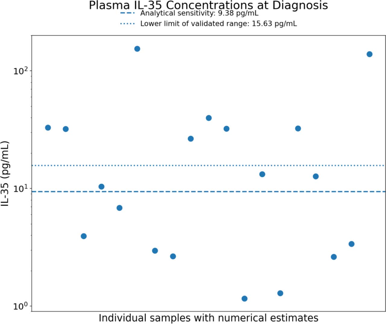
Figure 1.
Plasma IL-35 concentrations at diagnosis in mature T-cell neoplasms.
Individual numerical estimates are shown on a logarithmic scale. The dashed horizontal line indicates the manufacturer's analytical sensitivity (9.38 pg/mL), and the dotted horizontal line indicates the lower limit of the validated range (15.63 pg/mL). Numerical estimates below these thresholds are displayed to illustrate the observed distribution but should not be interpreted as equally precise quantitative measurements. The sample reported as below the detection limit is not shown.
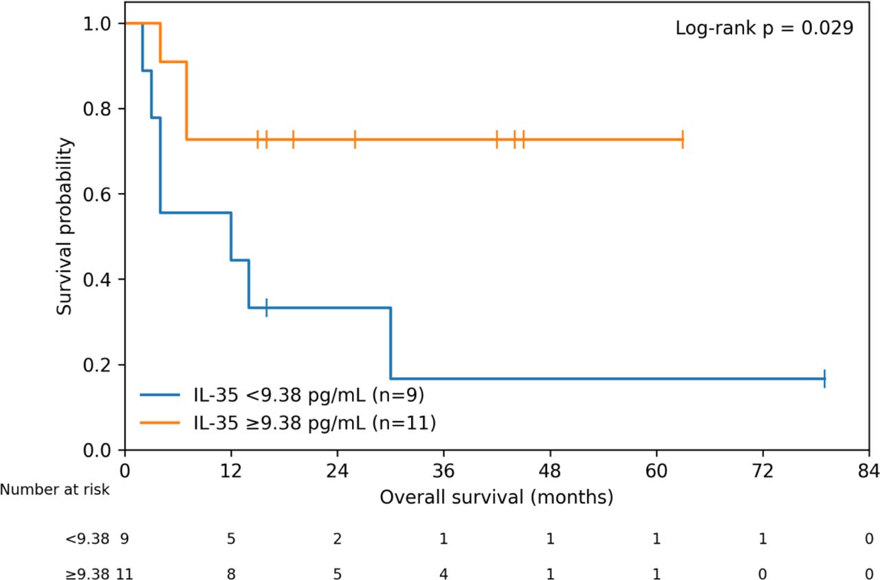
Figure 2.
Kaplan–Meier analysis of overall survival according to plasma IL-35 relative to the manufacturer's analytical sensitivity threshold. Patients were categorized as IL-35 ≥9.38 pg/mL (n = 11) or <9.38 pg/mL (n = 9). Higher IL-35 was associated with longer overall survival (log-rank p = 0.029; Cox HR 0.26, 95% CI 0.07–0.99; p = 0.049). Censored observations are indicated by tick marks. Numbers at risk are shown below the plot.
The result was consistent in a sensitivity analysis using 15.63 pg/mL, the lower boundary of the validated quantitative assay range. Eight patients had IL-35 values ≥15.63 pg/mL, of whom 1 died, compared with 9 deaths among 12 patients below 15.63 pg/mL. The ≥15.63 pg/mL group had longer OS (log-rank p = 0.012), with a univariable HR for death of 0.12 (95% CI 0.015–0.92; p = 0.041).
Because the overall cohort included two patients with primary cutaneous T-cell neoplasms (P15, CTCL; P17, mycosis fungoides with large-cell transformation), a disease scope sensitivity analysis was restricted to the 18 patients with systemic T-cell neoplasms. The association between higher IL-35 and longer OS was preserved. Using the primary analytical-sensitivity threshold of 9.38 pg/mL, IL-35 ≥9.38 pg/mL was associated with a lower hazard of death (HR 0.23, 95% CI 0.058–0.89; p = 0.033; log-rank p = 0.016). Using 15.63 pg/mL, the lower boundary of the validated quantitative range, the association remained evident (HR 0.076, 95% CI 0.009–0.61; p = 0.015; log-rank p = 0.0015). These disease-scope analyses were considered exploratory because of the small number of patients and events.
3.4. Plasma IL-2 and IL-10 Concentrations
IL-2 and IL-10 were evaluable in 11 patients and were quantifiable in only three patients each; because of the small number of measurable values, these cytokines were considered descriptive and were not subjected to formal survival analysis.
Table 3.
Descriptive distribution of disease entity, IPI category, and first-line treatment according to the primary IL-35 assay threshold.
| Characteristic | IL-35 <9.38 pg/mL (n=9) | IL-35 ≥9.38 pg/mL (n=11) |
|---|---|---|
| ALK-negative ALCL | 1 (11.1%) | 4 (36.4%) |
| PTCL-NOS | 1 (11.1%) | 1 (9.1%) |
| Nodal TFH lymphoma | 1 (11.1%) | 2 (18.2%) |
| ATLL | 3 (33.3%) | 2 (18.2%) |
| Intestinal T-cell lymphoma (EATL/MEITL) | 1 (11.1%) | 1 (9.1%) |
| ENKTL | 1 (11.1%) | 0 |
| Primary cutaneous T-cell neoplasm | 1 (11.1%) | 1 (9.1%) |
| IPI category Low | 5 (55.6%) | 5 (45.5%) |
| IPI category Low-intermediate | 1 (11.1%) | 2 (18.2%) |
| IPI category High-intermediate | 2 (22.2%) | 3 (27.3%) |
| IPI category High | 1 (11.1%) | 1 (9.1%) |
| First line treatment CHOEP | 4 (44.4%) | 3 (27.3%) |
| First line treatment BV-CHP | 0 | 3 (27.3%) |
| First line treatment CHOP | 1 (11.1%) | 1 (9.1%) |
| First line treatment CHOEP + HD-MTX | 0 | 1 (9.1%) |
| First line treatment PEG-IFN + zidovudine | 1 (11.1%) | 1 (9.1%) |
| Newcastle regimen | 0 | 1 (9.1%) |
| SMILE | 1 (11.1%) | 0 |
| Excision | 1 (11.1%) | 0 |
| Gemcitabine | 0 | 1 (9.1%) |
| GDP | 1 (11.1%) | 0 |
[i] Data are presented as n (% within IL-35 group). No inferential comparisons were performed. ATLL subtypes were grouped together; EATL and MEITL were grouped as intestinal T-cell lymphomas; the two primary cutaneous neoplasms were grouped together. BDL values were assigned to the <9.38 pg/mL group according to the assay-defined primary threshold.
4. DISCUSSION
The principal finding of this exploratory study was an association between plasma IL-35 above assay defined analytical thresholds and longer overall survival. The direction of the association was consistent using both the manufacturer's analytical sensitivity threshold and the lower boundary of the validated quantitative range, and it was preserved when the analysis was restricted to systemic T-cell neoplasms. The concordant direction of effect across the two assay defined thresholds and in the systemic disease sensitivity analysis supports the internal consistency of the observed association. Nevertheless, the small number of patients and events precludes conclusions regarding the independence or precise magnitude of the prognostic effect.
The association between higher IL-35 levels and longer survival is somewhat unexpected, given its generally immunosuppressive role. IL-35 is produced mainly by regulatory immune cells and can suppress effector T-cell responses and promote tumor immune escape [23,24,25,26,27,28]. However, immune regulation in lymphoma is more complex, and regulatory T-cell populations have not consistently been associated with poor outcome [29,30]. In our cohort, higher circulating IL-35 may therefore reflect a distinct immune environment rather than a direct protective effect of the cytokine. Since plasma measurements cannot identify the cellular source of IL-35 or reflect its activity within the tumor microenvironment, the biological basis of this association remains unclear.
IL-35 may also circulate in biologically distinct compartments, including extracellular vesicle associated forms, and experimental studies support a role for EV associated IL-35 signaling [33,34,35]. Because the present study measured total plasma IL-35 without isolating or characterizing the EV fraction, the relative contribution of soluble and EV-associated IL-35 to the observed survival association cannot be determined.
This study has several limitations. The cohort was small, histologically heterogeneous, and selected according to availability of stored diagnostic plasma rather than as a consecutive clinical series. The number of deaths was limited, precluding reliable multivariable or histology-specific modeling. Treatment exposure and treatment response were heterogeneous across the cohort and may have acted as important confounders of the observed association between IL-35 and overall survival; the limited sample size precluded reliable adjustment for these factors. In addition, many IL-35 estimates fell below the validated quantitative range of the assay, which is why the survival analyses were based on assay defined categorical thresholds rather than precise continuous low range concentrations. The EEL067 assay is a solid phase sandwich ELISA specifically designed and quality tested by the manufacturer for detection of human IL-35. The sandwich format requires sequential capture and detection of the target at different epitopes, with unbound components removed during washing steps. Previous methodological work using anti-p35 capture and anti-EBI3 detection demonstrated that a heterosubunit p35/EBI3 sandwich ELISA can specifically detect IL-35 without cross-reactivity with unpaired EBI3, IL-12, or IL-27 [36]. For EEL067, however, the precise proprietary epitopes recognized by the capture and detection antibodies are not publicly disclosed; therefore, we do not infer their individual subunit specificity beyond the manufacturer's validation of the assay for human IL-35. The inclusion of biologically distinct mature T-cell neoplasms introduces additional disease related heterogeneity; however, the direction of the IL-35 association was preserved in the sensitivity analysis restricted to systemic disease. Further subdivision into aggressive versus indolent categories was not considered sufficiently robust for inferential analysis in this sample, and larger disease-specific cohorts will be required for adjusted and subtype specific analyses.
5. CONCLUSION
In this exploratory cohort of mature T-cell neoplasms, higher plasma IL-35 relative to assay-defined analytical thresholds was associated with longer overall survival. The direction of this association was preserved using the lower boundary of the validated quantitative assay range and in a sensitivity analysis restricted to patients with systemic T-cell neoplasms. Given the small sample size, histological heterogeneity, limited number of events, and analytical constraints at low IL-35 concentrations, these findings should be regarded as hypothesis-generating rather than evidence of an independent prognostic effect. Larger, disease specific prospective studies using assays optimized for low range IL-35 quantification are required to determine whether plasma IL-35 has reproducible prognostic value.
Abbreviations
- AITL
angioimmunoblastic T-cell lymphoma
- ALCL
anaplastic large cell lymphoma
- ATLL
adult T-cell leukemia/lymphoma
- CTCL
cutaneous T-cell lymphoma
- EATL
enteropathy-associated T-cell lymphoma
- ELISA
enzyme-linked immunosorbent assay
- ENKTL
extranodal NK/T-cell lymphoma
- EV
extracellular vesicle
- IPI
International Prognostic Index
- IL
interleukin
- MEITL
monomorphic epitheliotropic intestinal T-cell lymphoma
- nTFHL
nodal T-follicular helper cell lymphoma
- OS
overall survival
- PTCL
peripheral T-cell lymphoma
- Treg
regulatory T cell.
Notes
[5] Ethics Approval:
The patient-derived biological samples used in this study were collected at the Department of Hematology, Fundeni Clinical Institute, in accordance with institutional ethical requirements and with the approval of the Institutional Ethics Committee in 2019 (Approval No. 3807/02.08.2019). The additional biomarker analyses and retrospective evaluation of associated clinical data performed for the present study were subsequently approved by the Ethics Committee of Fundeni Clinical Institute (approval No. 30164, 18 July 2025).
[7] Data Availability:
Further data are available from the corresponding author on reasonable request, subject to applicable ethical and data-protection requirements.
[9] Authorship
Conceptualization: A.B., M.U.; Methodology: A.B., M.U., C.G.; Investigation: A.B., M.U., C.G.; Data curation: A.B., M.U.; Formal analysis: A.B.; Writing – original draft: A.B.; Writing – review and editing: M.U., C.G., D.C.; Validation: D.C.; Supervision: D.C.; Resources and clinical support: A.M.C., V.T., M.E.T., A.Bu., S.H.M., C.D.; Data collection and clinical characterization: A.M.C., V.T., M.E.T., A.Bu., S.H.M., C.D. All authors reviewed and approved the final version of the manuscript.