
Introduction
Primary angiitis of the central nervous system (PACNS) is an extremely rare diagnosis, with an incidence of 2.4 cases per 1,000,000 person-years[1]. The exact cause of PACNS remains unknown, and typically cases present with a long prodrome. It is a diagnosis of exclusion, and various infectious, alternative autoimmune, and malignant processes must be excluded prior to final diagnosis[2]. Given the rarity of this condition there is often a delay in diagnosis that can range from 6 to 23 months [3,4]. We present a case of PACNS that responded well to combination therapy with high-dose steroids and cyclophosphamide.
Case Presentation
A 70-year-old Caucasian male with history of hyperlipidemia, hypertension, former smoker (12 pack years), benign prostate hyperplasia, obesity and remote history of left ophthalmic stroke 4 years prior presented to the emergency department (ED) for a 3-week history of unintentional weight loss of 10 pounds, headache, decreased appetite, nausea, weakness, fatigue, and night sweats. He denied burning chest pain, dysphagia, odynophagia, epigastric pain, abdominal pain, stool changes, joint pain, dyspnea, numbness/tingling or new rash. Family history was significant for a father with diabetes mellitus and heart disease, and a mother with lung cancer. Social history was significant for alcohol use 3–5 times per week. Physical exam was grossly unremarkable apart from left eye horizontal diplopia. Specifically, neurological exam was notable for left eye horizontal diplopia, intact sensation to light touch, 5/5 motor strength with no drift in the arms and legs bilaterally, and 2+ deep tendon reflexes throughout. Of note, the patient had a magnetic resonance image (MRI) with and without contrast of the brain at an outside hospital that was reported as unremarkable.
Initial workup in the ED was significant for mild anemia and mild leukocytosis of 11.6 × 10⁹/L. C-reactive protein (CRP) was mildly elevated at 11.2 mg/L, and erythrocyte sedimentation rate was within normal limits. Parathyroid hormone was normal, morning cortisol was within normal limits, LDH 130 U/L (normal), angiotensin converting enzyme level 33 nmol/mL/min (normal), adrenocorticotropic hormone 15 pg/mL (normal), thyroid stimulating hormone 2.8 mU/L (normal), free thyroxine 1.7 ng/dL (normal), antinuclear antibody 0.8 IU/mL (within normal limits), and anti-cyclic citrullinated peptide antibody <15.6 U/mL (normal). C-ANCA negative, P-ANCA negative, beta-2 GPR IgG Ab <9.4 U/mL (negative), anti-phospholipid antibody IgG <9.4 U/mL (negative), anti-double stranded DNA antibody <12.3 U/mL (negative), anti-SSA/SSB IgG antibody <0.2 U/mL (negative), anti-Smith antibody IgG less than 0.2 /mL (negative), serum rheumatoid factor <15 U/mL (normal), anti-centromere antibody IgG less than 0.2 U/mL (negative), anti-Scl 70 antibody IgG less than 0.2 U/mL (negative), anti-Jo 1 antibody IgG less than 0.2 U/mL (negative), and anti-myeloperoxidase antibody less than 0.2 U/mL (negative). There was no apparent monoclonal protein on serum electrophoresis. Computed tomography (CT) of the abdomen and pelvis with intravenous (IV) contrast was unrevealing apart from focal soft tissue thickening with calcification in the medial left lung base. The patient was referred to gastroenterology for further assessment of possible primary gastrointestinal malignancy in the setting of mild anemia of unknown duration and unexplained weight loss. Upper endoscopy found inactive chronic gastritis and duodenal erosions without bleeding with nonspecific reactive changes, and there was no malabsorption pattern. Colonoscopy removed a 3 mm sessile hyperplastic polyp from the ascending colon and identified diverticulosis. A chest CT scan was ordered given prior abnormal lung findings which showed two 3 mm pulmonary nodules in the right middle lobe and the left lower lobe.
Repeat brain MRI with and without IV contrast showed numerous new small focal and ill-defined patchy enhancing lesions on T2 hyperintensity located throughout both cerebral hemispheres. A few lesions were also located in both cerebellar hemispheres. There was no mass effect, surrounding edema, hemorrhage or restricted diffusion associated with these lesions. Overall, the lesions were nonspecific. Differential diagnosis included but was not limited to diffuse parenchymal and leptomeningeal metastases, primary CNS lymphoma, Moya Moya angiopathy, neurosarcoidosis, PACNS, and CNS vasculitis secondary to possible viral, bacterial, or fungal causes. There was no evidence of intracranial abscess.
Lumbar puncture revealed clear liquid with 65 nucleated cells/microliter, <1 erythrocyte/microliter, 5 neutrophils/microliter, 85 lymphocytes/microliter, and no eosinophils, basophils, or other cells. Total protein 82 mg/dL, glucose 54 mg/dL, elevated B2 microglobulin 2.41 mcg/mL, and negative Gram stain. Serum glucose obtained near the time of lumbar puncture was 100 mg/dL, and the CSF to serum glucose ratio was normal at 0.54. Therefore, hypoglycorrhachia was not present. Cerebrospinal fluid bacterial (aerobic and anaerobic) cultures negative, mycobacterial cultures negative, fungal cultures negative, negative viral panel, and negative panel for malignancy.
One month after initial presentation, the patient was admitted to the hospital for reported monocular horizontal diplopia of the left eye, worsening short term memory, slurred speech, gait imbalance, weakness, fatigue, somnolence, bifrontal headaches and 20-pound weight loss over a 2-week period. The patient and his wife also noted that over the past couple of weeks he had experienced intermittent numbness and tingling of his left jaw. Patient underwent ophthalmology evaluation in the outpatient setting which did not reveal a specific structural cause of monocular horizontal diplopia and noted no papilledema. In the next few weeks, his reported monocular horizontal diplopia of his left eye resolved. MRI cervical and thoracic spine did not reveal bony metastatic disease in these regions but revealed 2 tiny foci of leptomeningeal enhancement which were indeterminate in the thoracic cord dorsal surface at T10 and T11 levels.
Whole body fluorodeoxyglucose F 18 positron emission tomography/computed tomography was pursued as a screen for an unknown occult malignancy. It demonstrated intense focal abnormal increased uptake in the inferior right cerebellum which was concerning for an infectious, inflammatory, or neoplastic process. The patient was followed in the outpatient setting by neurology and initially experienced clinical improvement without targeted therapy.
Three months later, the patient exhibited progressive cognitive decline, especially short-term memory loss, decreased executive function and imbalanced gait, further episodes of monocular horizontal diplopia and continued weight loss of 10 additional pounds. The patient was admitted for further work-up of worsened symptoms. Further workup revealed a negative CSF paraneoplastic panel (negative histopathological and immunohistochemical findings) apart from a positive acetylcholine receptor ganglionic neuronal antibody (0.13 nmol/L) and a borderline rheumatoid factor (RF) of 17 IU/mL.
Repeat MRI of the brain showed diffuse, multiple new enhancing lesions with resolution of previously seen lesions in mild diffusion restriction in the right paracentral region (Figure 1). MRA of the brain demonstrated diffuse signal loss involving the intracranial portion of the right vertebral artery.
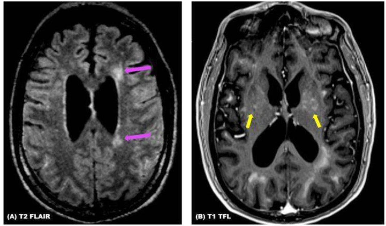
Figure 1
(A and B) Stealth MRI brain protocol demonstrates the presence of scattered patchy and confluent foci of FLAIR hyperintensity and abnormal subcortical white matter enhancement predominantly in cerebral hemispheres (arrows in (A)), but also involving the corpus callosum, basal ganglia bilaterally (arrows in (B)), the right anteromedial thalamus, middle cerebellar peduncles, subcortical white matter in the cerebellar hemispheres and the medulla. These findings were suggestive of Primary Angiitis of the Central Nervous System (PACNS) in the absence of an underlying systemic vasculitis.
As noted by the radiologist, the MRI and MRA brain findings were concerning for a large vessel vasculitis. A brain biopsy was pursued for definitive diagnosis. Brain biopsy demonstrated perivascular and parenchymal lymphohistiocytic infiltrates with small vessel vasculitis, endothelial swelling, and accumulation of pigmented macrophages and eosinophils around damaged vessels. No noncaseating granulomas were identified. There was a small vessel vasculitis with inflammation, endothelial swelling, and accumulation of pigmented macrophages and eosinophils around damaged vessels. Bacterial (aerobic and anaerobic), mycobacterial, and fungal cultures from the brain biopsy were negative. Cytomegalovirus polymerase chain reaction was negative. In the context of the patient’s negative prior alternative systemic autoimmune work-up, negative HIV, syphillis, and Borrelia burgdorferi infectious work-up, and negative malignancy work-up, the most likely diagnosis was deemed PACNS.
The patient was treated with empiric high-dose methylprednisolone 1000 mg for 5 days and was referred to rheumatology for further management. The patient was started on monthly IV cyclophosphamide infusions 500 mg/m2 body surface area and prednisone 60 mg daily with planned taper. He tolerated cyclophosphamide well, with subsequent labs demonstrating no cytopenia and no concern for hepatotoxicity. Overall, the patient did not worsen from a neurologic standpoint, but his baseline neurologic deficits persisted.
Discussion
PACNS is an extremely rare disease and remains a challenge to recognize clinically. It is a diagnosis of exclusion, given that its non-specific imaging and histopathology findings share similar features with other autoimmune, infectious, and malignant diseases [2]. MRI of the brain with and without contrast commonly demonstrates multiple strokes in multiple vascular territories [5]. Histopathology of PACNS classically demonstrates the presence of Langerhans or foreign body giant cells, necrotizing vasculitis, or lymphocytic vasculitis [6]. The diagnosis of PACNS is confirmed if the patient has an acquired unexplained neurological deficit, has the classic imaging and histopathology findings, and has no evidence of another autoimmune, infectious, or malignant disease that could mimic the imaging and histopathology findings [6,7].
It is imperative to ensure a negative infectious work-up when considering PACNS for two reasons. First, there are several documented subacute and chronic infectious processes that are known to mimic PACNS, with causative organisms including but not limited to Treponema pallidum (neurosyphilis), Borrelia burgdorferi (Lyme disease), Mycobacterium tuberculosis, hepatitis B and C viruses, HIV, Varicella-Zoster virus, and Aspergillus, Coccidioides, and Histoplasma species [2]. Second, the treatment for PACNS requires potent immunosuppressive therapy, and if an underlying infectious process is present, immunosuppressive therapy can cause rapid clinical deterioration. Our patient had an extensive infectious work-up testing of both the CSF as well as the brain biopsy which was pan-negative.
Additionally, it is important to exclude alternative autoimmune causes of CNS dysfunction, as PACNS is a diagnosis of exclusion. This patient presented with a mildly elevated RF level as well as the positive acetylcholine receptor ganglionic neuronal antibody. Rheumatoid meningoencephalitis was deemed less likely given the lack of additional abnormal autoimmune testing and articular/extraarticular rheumatoid arthritis manifestations in the patient [8].
A challenging aspect of this case was the interpretation of neuroimaging. Specifically, the MRI brain with and without contrast and MRA brain findings were indicative of a possible large vessel vasculitis, since the intracranial portion of the right vertebral artery demonstrated diffuse signal loss. Given these findings, it has been proposed in the literature that digital subtraction angiography (DSA) should be pursued instead of brain biopsy when considering the diagnosis of PACNS because vasculitis of medium and large vessels may not be detected in a brain biopsy sample [2,7]. Furthermore, a systematic review and meta-analysis of 46 cohort studies assessing 911 PACNS patients demonstrated that DSA was positive in 33% of brain biopsy-confirmed cases of PACNS, and brain biopsy was positive in only 8% of DSA-confirmed cases of PACNS [9]. The low overlap between brain biopsy and DSA suggests different histopathologic types of PACNS, with the caveat being that it was unclear from the study methods if all cases with brain biopsy also had DSA [9]. Additionally, there are 2 significant mimickers of PACNS, Moya Moya angiopathy and reversible cerebral vasoconstriction syndrome (RCVS). DSA remains the gold standard for definitive diagnosis of Moya Moya angiopathy, and DSA with concomitant intraarterial nimodipine can confirm RCVS [2]. Although DSA was not performed, the overall clinical presentation was less consistent with RCVS or Moya Moya angiopathy given the inflammatory CSF profile, leptomeningeal enhancement, progressive neurologic decline, constitutional symptoms, and brain biopsy demonstrating inflammatory vasculitic changes.
Neurosarcoidosis was also carefully considered in the differential diagnosis given the patient’s constitutional symptoms, weight loss, mild inflammatory marker elevation, leptomeningeal enhancement, pulmonary nodules, and responsiveness to corticosteroids. Additionally, the histopathologic findings of lymphohistiocytic inflammation may overlap with reported findings in neurosarcoidosis. However, several features favored PACNS over neurosarcoidosis in this case. The patient lacked more characteristic systemic manifestations of sarcoidosis including hilar or mediastinal lymphadenopathy, cutaneous disease, ocular inflammation, hypercalcemia, or other organ involvement. Pulmonary imaging demonstrated only two small nonspecific pulmonary nodules and focal calcification without classic thoracic imaging findings suggestive of sarcoidosis. Furthermore, serum angiotensin converting enzyme level was normal, although we acknowledge that this finding alone does not exclude sarcoidosis, particularly extrapulmonary disease. Brain biopsy did not demonstrate noncaseating granulomas, and infectious, malignant, and alternative systemic autoimmune evaluations were unrevealing. Taken together, the overall clinical, radiographic, and histopathologic presentation was felt to be most consistent with PACNS, although neurosarcoidosis could not be definitively excluded.
There is frequently delay in the diagnosis of PACNS. A retrospective study of 28 biopsy-proven cases of PACNS demonstrated a mean time to diagnosis of 6 months [3]. However, a different observational study of 82 confirmed cases of PACNS had a median time to diagnosis of 23 months, highlighting the rarity and/or lack of initial awareness of this diagnosis at the initial time of presentation among all clinical providers [4].
Management of PACNS includes combination therapy with glucocorticoids and cyclophosphamide [10,11,12]. Patients are typically managed with high-dose prednisone 1 mg/kg (max 80 mg/day) or started on methylprednisolone 15 mg/kg for 3 days (max 1 g) with transition to oral prednisone on day 4 [12]. IV cyclophosphamide is typically started at 600-750 mg/m2, with dose reductions for patients with kidney dysfunction [13,14]. However, it should be noted that this cyclophosphamide dosing is based off dosing for treatment of systemic vasculitis, and PACNS is not considered a systemic vasculitis. Our patient was started on high-intensity methylprednisolone 1000 mg per day for 5 days in the hospital followed by prednisone 60 mg daily as indicated by rheumatology. The patient was treated with IV cyclophosphamide 500 mg monthly in the absence of kidney dysfunction, which he tolerated well. There is a lack of randomized control trial data to confirm that higher doses of cyclophosphamide are associated with lower mortality or reduced disease recurrence versus lower doses of cyclophosphamide for PACNS patients. Failure to respond to empiric cyclophosphamide should prompt additional evaluation for an alternative diagnosis before escalating treatment or trialing another immunosuppressive agent. In patients who cannot receive cyclophosphamide due to underlying comorbidities or intolerances, Rituximab infusions (either as two 1 gram infusions separated by 14 days or as 375 mg/m2 weekly for four weeks) may be used. Three reported cases describe clinical and radiographic improvement in adults with PACNS following treatment with rituximab [15,16]. In one case, PACNS was diagnosed based on angiographic findings and abnormal cerebrospinal fluid (CSF) analysis, and rituximab was used as first-line therapy [15]. In another case, the diagnosis was pathologically confirmed by evidence of a lymphocytic vasculitic process involving the brain and spinal cord; rituximab was initiated after the patient failed to improve following five monthly cyclophosphamide infusions [15]. In the third case, pathologic examination demonstrated granulomatous vasculitis, and rituximab was administered as initial therapy [16].
Finally, it is important to emphasize the importance of multi-disciplinary nature of this case. The patient received care from internal medicine physicians, neurologists, neuroradiologists, and rheumatologists to aid in diagnosis of this condition. There were several month-long delays in diagnosis with multiple hospitalizations during that time span and therapy was thus delayed due to transient spontaneous clinical improvement. Moving forward, clinicians should remain mindful of the atypical and often non-specific clinical presentations and imaging findings associated with PACNS when developing their differential diagnoses, as delayed or missed recognition can lead to devastating consequences for the patient.
Conclusions
PACNS is an extremely rare disease with a non-specific clinical presentation and has devastating long-term consequences if diagnosis is delayed. All infectious, alternative autoimmune, and malignant mimicking processes must be excluded prior to diagnosis of PACNS. If imaging findings are indicative of medium or large vessel vasculitis and RCVS versus moyamoya angiopathy is high on the differential, clinical providers should consider DSA initially over brain biopsy. PACNS requires multi-disciplinary assistance in regard to both diagnosis and management from internal medicine physicians, rheumatologists, neuroradiologists and neurologists. Delays in diagnosis of PACNS are common, with multiple studies reporting delays in diagnosis ranging from 6–23 months. Once the diagnosis is confirmed, management includes combination therapy typically with high-dose steroids and cyclophosphamide, although this is based off observational study data, case reports, and case series. Randomized controlled trials are needed to establish optimal dosing efficacy for reduced mortality and reduced disease recurrence for patients with PACNS.
Limitations
A limitation of this case was the inability to definitively exclude neurosarcoidosis, particularly given the constitutional symptoms and nonspecific pulmonary findings. Soluble IL-2 receptor testing may have provided additional diagnostic utility given its reported sensitivity of 88% and specificity of 87% in extrapulmonary sarcoidosis, although this was not obtained during the patient’s evaluation [17]. An additional limitation of this case was the lack of definitive understanding of the mechanism of monocular diplopia.
Acknowledgements
The authors have no acknowledgements to add.