
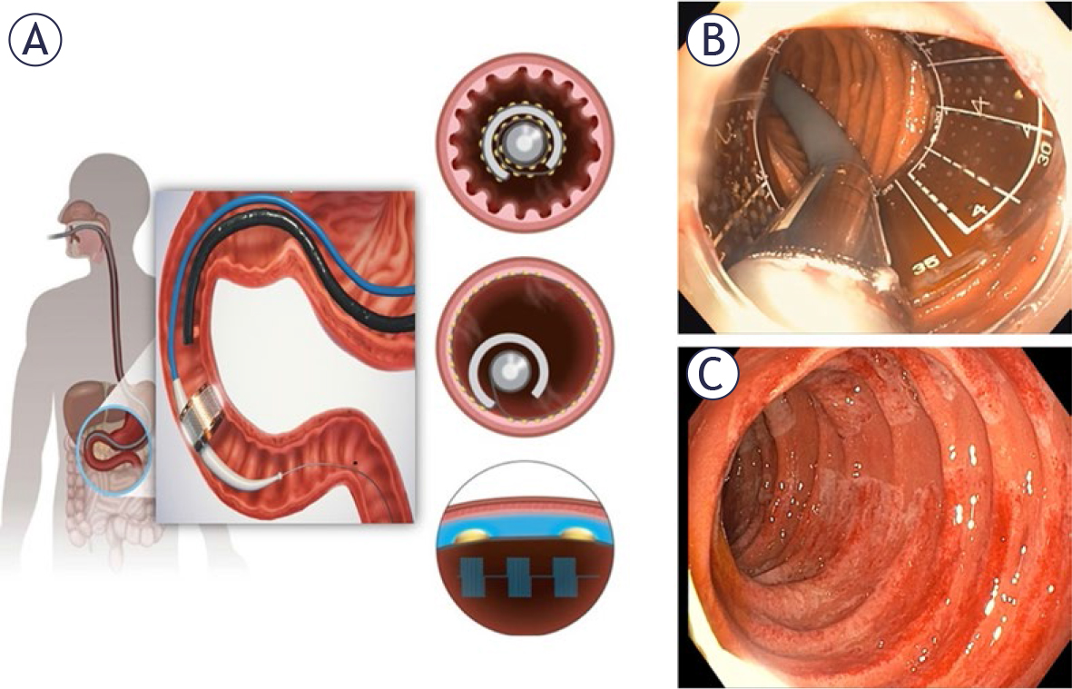
Figure 1.
Recellularization via electroporation therapy (ReCET) is a novel endoscopic procedure that uses an endoscope-mounted electrode to deliver pulsed electric fields to the duodenal mucosa and submucosa. (A) A schematic of the positioning of the flexible electrode and its unfolding and expansion within the duodenum. (B) Intraprocedural endoscopic view of the expanded electrode within the duodenum. (C) A post-treatment endoscopic view of the resulting circumferential, nonthermal treatment effect. Figure is reproduced from.62
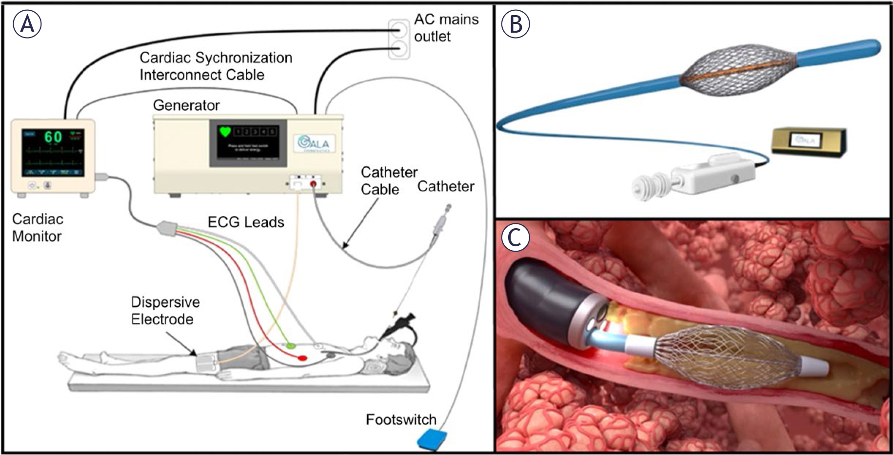
FIGURE 2.
(A) The RheOx system (Galvanize Therapeutics, Redwood City, CA, USA) for the treatment of the symptoms of chronic bronchitis consists of a proprietary electrosurgical generator and (B) a single use endoscope with an expandable basket electrode. (C) The minimally invasive bronchoscopic therapy delivers short bursts of Pulsed Electric Field (PEF) energy to induce non-thermal ablation of the abnormal mucus-producing cells in the airways, leaving the extracellular matrix intact so that the epithelium may rapidly regenerate. Figure is reproduced from.66
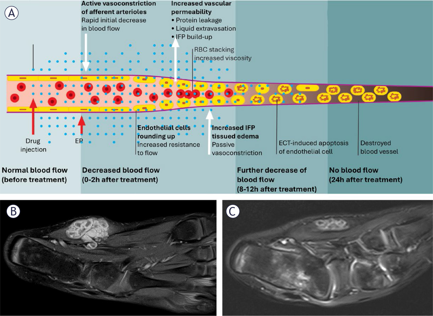
FIGURE 3.
Bleomycin ElectroScleroTherapy (BEST) combines intralesional bleomycin with reversible electroporation, exploiting the same vascular-disrupting mechanism described for electrochemotherapy (ECT). (A) An illustration of blood-flow changes induced by electrochemotherapy (ECT) at the level of a microcirculatory vessel; from normal flow before treatment to vascular destruction by 24 h. Adapted from.12 An example of BEST treatment of a vascular malformation in the foot: (B) T2-weighted MRI of a vascular malformation in the foot before BEST treatment, showing the hyperintense malformation. (C) MRI of the same region 3 months after BEST, demonstrating a 75% volume reduction.
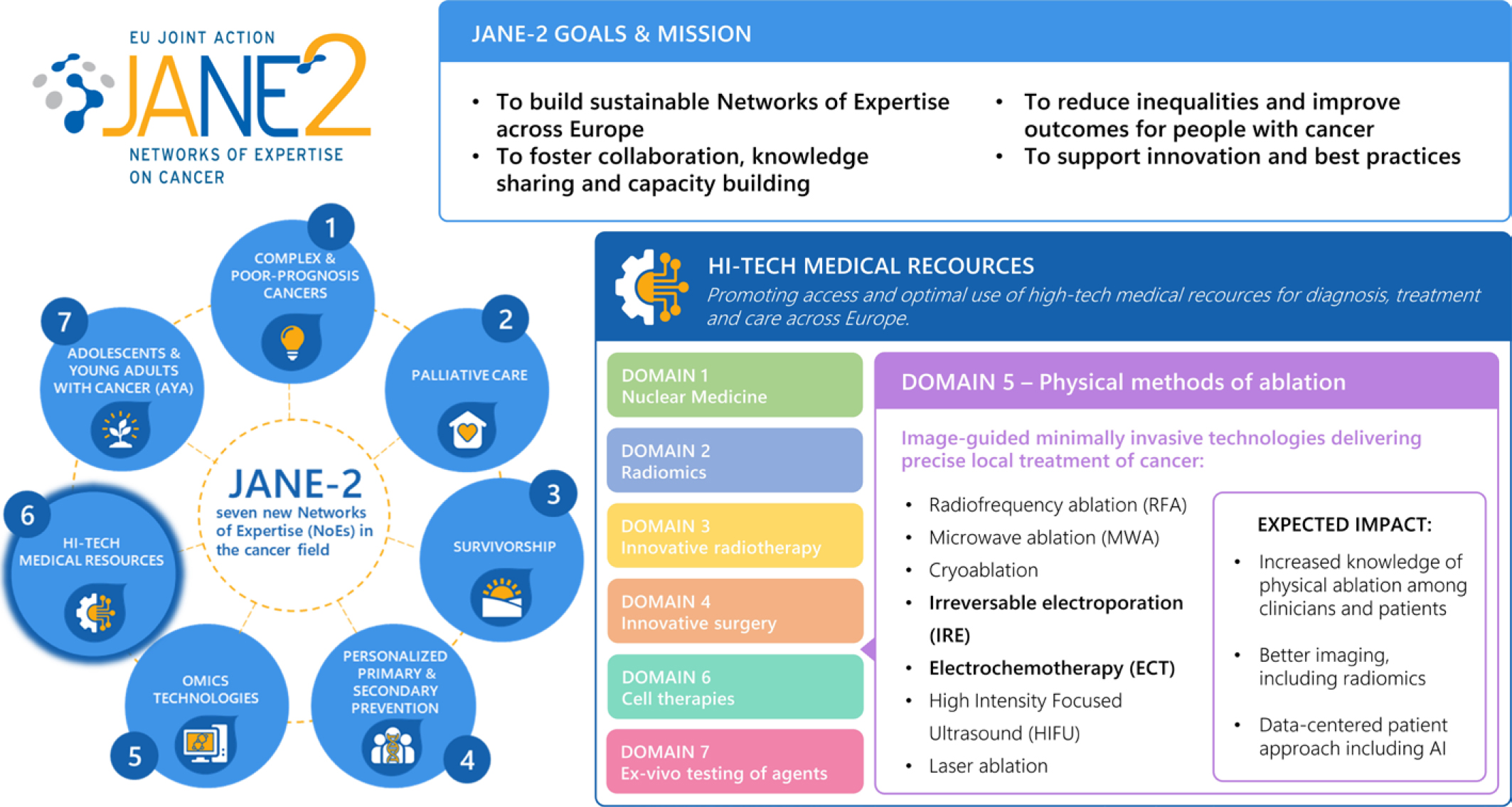
FIGURE 4.
Overview of the JANE-2 (Joint Action on Networks of Expertise on Cancer) framework, highlighting WP10 – High-Tech Medical Resources and Domain 5 - Physical Methods of Ablation. The figure illustrates the position of Domain 5 within the broader network of JANE-2 and emphasizes its expected impact on advancing equitable access to innovative cancer treatment technologies across Europe.
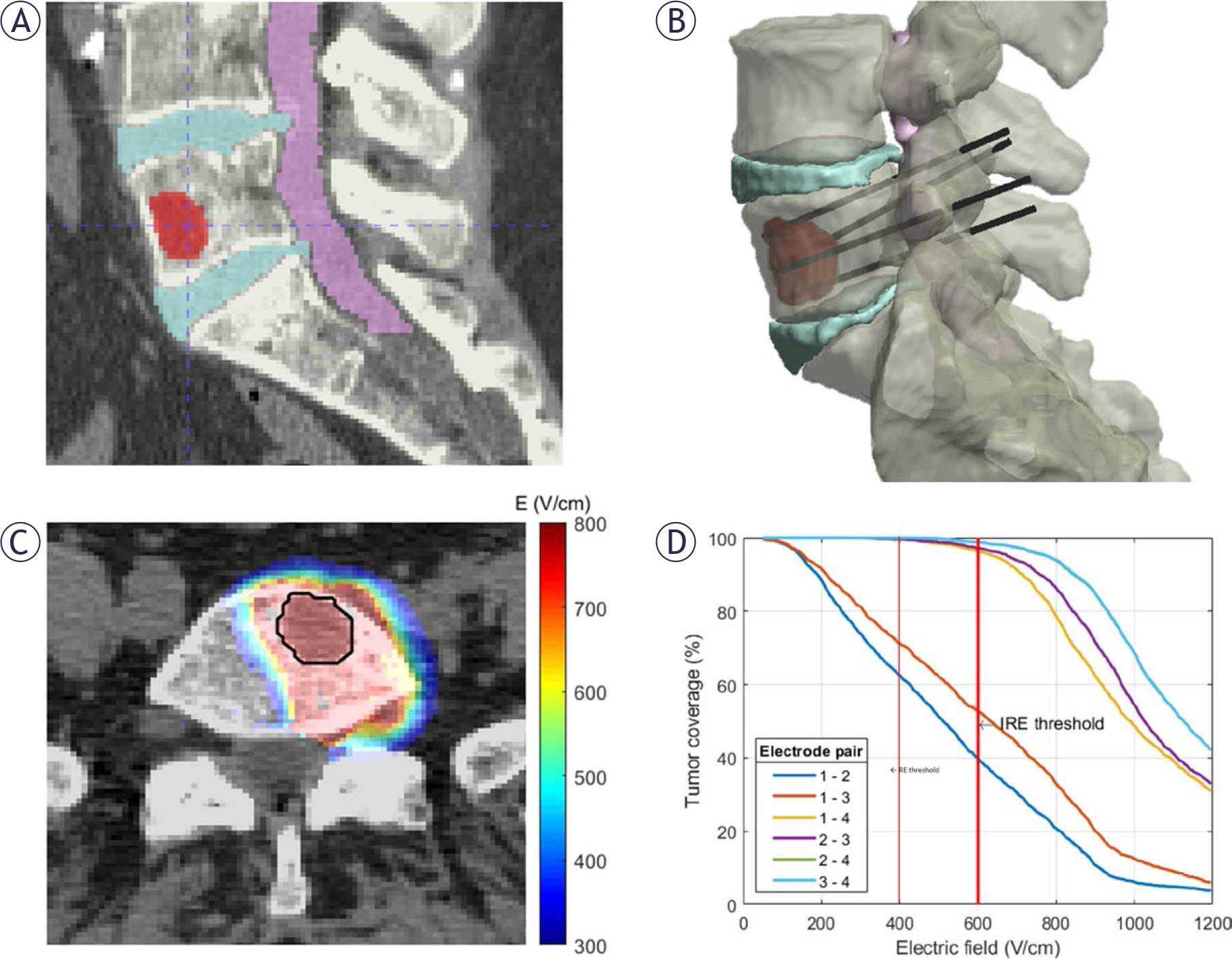
FIGURE 5.
Illustration of a computer-assisted patient-specific treatment plan for electrochemotherapy of a spinal metastasis. (A) Segmentation of the tissues of interest (vertebrae, intervertebral discs, spinal cord, tumour) from the patient’s CT image. (B) Patient-specific 3D model developed from image segmentation showing the electrode placement for the selected treatment plan. (C) Predicted electric field distribution for the selected treatment plan, overlaid on the CT image (tumour outlined in black). (D) Cumulative tumour coverage as a function of electric field for the candidate electrode pairs; vertical lines mark the reversible (RE) and irreversible (IRE) electroporation thresholds. Adapted from.106