
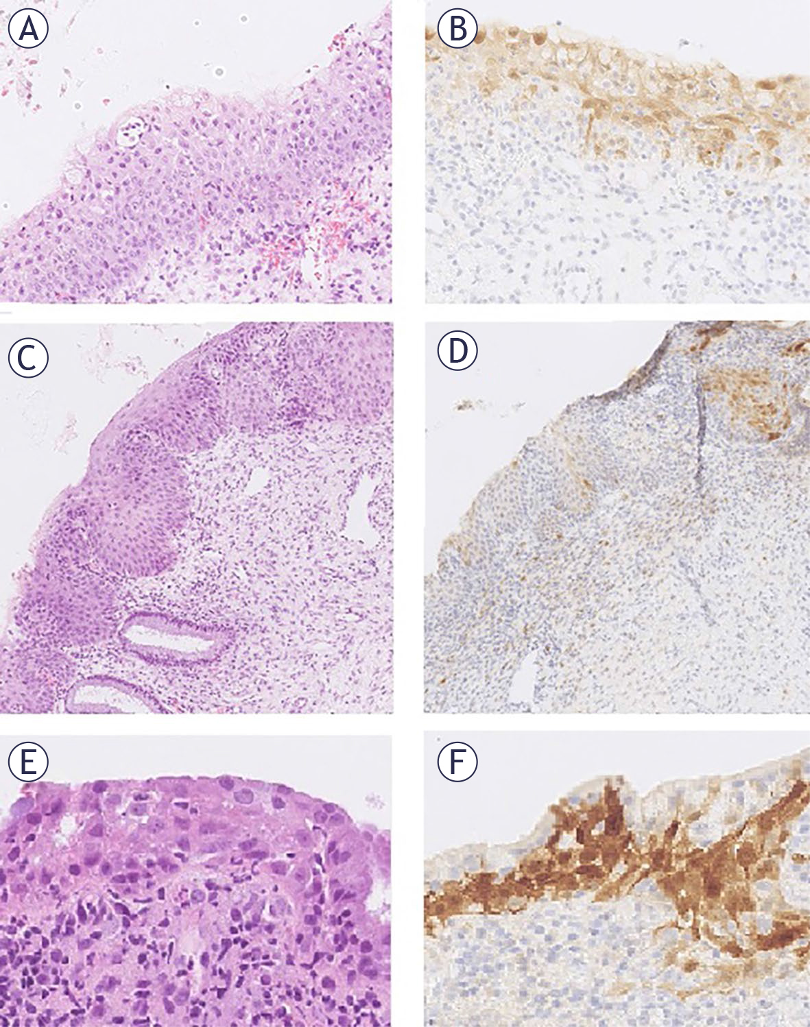
FIGURE 1.
Images of atypical squamous epithelium cases with patchy p16 reaction: (A–F). The same sample stained with HE and p16 at 200× magnification. Panels (A), (C), and (E) show HE staining, while panels (B), (D), and (F) show corresponding p16 staining.
TABLE 1.
Immunohistochemical reactions to various biological markers and human papillomavirus (HPV) genotyping in tissue biopsy and previous cervical smear, where available, by study group
| Group | p16 | N | Ki67 | N | E4 HPV | N | Nanog | N | HPV genotyping | N tissue | N smear |
|---|---|---|---|---|---|---|---|---|---|---|---|
| p16 null N = 11 | Null | 11 | 0 | 5 | Neg. | 11 | Neg. | 7 | Neg | 9 | 3 |
| Patchy | 0 | 1/3 | 4 | Pos. | 0 | 1 | 4 | lrHPV only | 2 | 1 | |
| En Block | 0 | 2/3 | 0 | 2 | 0 | hrHPV | 0 | 3 | |||
| 3/3 | 2 | 3 | 0 | ||||||||
| p16 patchy N = 24 | Null | 0 | 0 | 12 | Neg. | 23 | Neg. | 13 | Neg | 10 | 2 |
| Patchy | 24 | 1/3 | 5 | Pos. | 1 | 1 | 11 | lrHPV only | 6 | 2 | |
| En Block | 0 | 2/3 | 3 | 2 | 0 | hrHPV | 8 | 12 | |||
| 3/3 | 4 | 3 | 0 | ||||||||
| Condyloma N = 9 | Null | 0 | 0 | 0 | Neg. | 7 | Neg. | 0 | Neg | 0 | 1 |
| Patchy | 9 | 1/3 | 2 | Pos. | 2 | 1 | 9 | lrHPV only | 6 | 2 | |
| En Block | 0 | 2/3 | 1 | 2 | 0 | hrHPV | 3 | 1 | |||
| 3/3 | 6 | 3 | 0 | ||||||||
| CIN1 N = 12 | Null | 0 | 0 | 2 | Neg. | 2 | Neg. | 0 | Neg | 0 | 0 |
| Patchy | 10 | 1/3 | 2 | Pos. | 10 | 1 | 12 | lrHPV only | 6 | 1 | |
| En Block | 2 | 2/3 | 4 | 2 | 0 | hrHPV | 6 | 3 | |||
| 3/3 | 4 | 3 | 0 | ||||||||
| CIN2 N = 11 | Null | 0 | 0 | 0 | Neg. | 3 | Neg. | 0 | Neg | 1 | 0 |
| Patchy | 0 | 1/3 | 0 | Pos. | 8 | 1 | 7 | lrHPV only | 2 | 0 | |
| En Block | 11 | 2/3 | 2 | 2 | 4 | hrHPV | 8 | 3 | |||
| 3/3 | 9 | 3 | 0 | ||||||||
| CIN3 N = 9 | Null | 0 | 0 | 0 | Neg. | 7 | Neg. | 1 | Neg | 0 | 0 |
| Patchy | 0 | 1/3 | 0 | Pos. | 2 | 1 | 3 | lrHPV only | 0 | 0 | |
| En Block | 9 | 2/3 | 0 | 2 | 3 | hrHPV | 9 | 4 | |||
| 3/3 | 9 | 3 | 2 |
1 0 = null or parabasal reaction of Ki67; 1/3 = Ki67 positive in predominantly lower 1/3 of epithelium; 2/3 = Ki67 positive in predominantly lower 2/3 of epithelium; 3/3 = Ki67 positive in full thickness of epithelium; CIN = cervical intraepithelial neoplasia; hrHPV = high risk HPV; lrHPV = low risk HPV; Neg. = negative reaction; Pos. = positive reaction
TABLE 2.
Follow-up histology, cytology, human papillomavirus (HPV) result by study groups (p16 null and p16 patchy)
| Group | Follow up histo | N | Follow up cyto | N | Follow up HPV | N |
|---|---|---|---|---|---|---|
| p16 null (11 cases, 9 biopsies) | Neg | 0 | Neg | 5 | Neg | 5 |
| CIN1 | 1 | ASC-US | 4 | Pos | 3 | |
| CIN2 | 1 | LSIL | 0 | |||
| CIN3 | 1 | HSIL | 0 | |||
| p16 patchy (24 cases, 17 biopsies) | Neg | 0 | Neg | 5 | Neg | 4 |
| CIN1 | 2 | ASC-US | 6 | Pos | 5 | |
| CIN2 | 0 | LSIL | 4 | |||
| CIN3 | 3 | HSIL | 2 |
1 ASC-H = atypical squamous cells, cannot exclude HSIL; ASC-US = atypical squamous cells of undetermined significance; CIN = cervical intraepithelial neoplasia; Follow up cyto = cervical smears from follow-up; Follow up histo = histological samples from follow-up; Follow-up HPV = HPV results using the Hybrid Capture II assay; HSIL = highgrade squamous intraepithelial lesions; LSIL = low-grade squamous intraepithelial lesions; Neg = negative; Pos = positive
1 Follow-up after radical resections (conizations, cervical amputations, hysterectomies) was not informative, as the lesion was completely removed; therefore, this data is not included. In the case of multiple consecutive examinations (cytology, HPV testing), we only considered the most pathological cytology results or the positive HPV test.
TABLE 3.
Biomarkers in the primary biopsy and follow-up in the groups with atypical squamous epithelium with a patchy and a null reaction to p16
| Group | Case | Ki67 | E4 HPV | Nanog | HPV genotypes | Follow-up sample | Follow-up diagnosis | Time from primary biopsy to follow-up sample |
|---|---|---|---|---|---|---|---|---|
| p16 | 1 | 1/3 | - | 1+ | 31, 52 | biopsy | CIN1 | 2 years |
| null | 2 | 3/3 | - | - | 53 | biopsy | CIN2 | 2 years |
| 3 | 1/3 | - | - | - | biopsy | CIN3 | 4 months | |
| p16 patchy | 4 | 0 | - | - | 39 | cone | CIN1 | 8 months |
| 5 | 1/3 | - | - | - | cone | CIN1 | 6 years | |
| 6 | 3/3 | - | 1+ | 53 | cone | CIN3 | 5 years | |
| 7 | 3/3 | - | - | 52, 53, 56, 66, 73 | Cervical amputation | CIN3 | 10 months | |
| 8 | 1/3 | - | - | 44 | cone | CIN3 | 10 months |
1 0 = null or parabasal reaction of Ki67; 1/3 = Ki67 positive in predominantly lower 1/3 of epithelium; 2/3 = Ki67 positive in predominantly lower 2/3 of epithelium; 3/3 = Ki67 positive in full thickness of epithelium; - = negative reaction, + = positive reaction; CIN = cervical intraepithelial neoplasia; HPV = human papillomavirus
TABLE 4.
Comparison of different biological markers between groups using Spearman correlation
| Groups | p16 | Ki67 | HPV E4 | Nanog | lrHPV tissue | hrHPV tissue | hrHPV: tissue+smear | lrHPV: tissue+smear |
|---|---|---|---|---|---|---|---|---|
| p16 null vs. p16 patchy | r = 1 p = 0.000 | r = 0.000 | r = 0.116 | r = 0.089 | r = 0.209 | r = 0.384 p = 0.025 | r = 0.274 | r = 0.285 |
| p16 patchy vs. condyloma | r = 0.000 | r = 0.535 p = 0.001 | r =0.280 | r = 0.494 p = 0.004 | r = 0.458 p = 0.007 | r = 0.000 | r=-0.186 | r = 0.494 p = 0.004 |
| p16 patchy vs. CIN1 | r = 0343 p = 0.041 | r = 0.35 p = 0.037 | r = 0.81 p = 0.000 | r = 0.532 p = 0.001 | r = 0.354 p = 0.034 | r = 0.161 | r = -0.039 | r = 0.359 p = 0.032 |
| p16 patchy vs. CIN2 | r = 1 p = 0.000 | r = 0.66 p = 0.000 | r = 0.728 p = 0.000 | r = 0.632 p = 0.000 | r = -0.193 | r = 0.45 p = 0.007 | r = 0.176 | r = -0.089 |
| p16 patchy vs. CIN3 | r = 1 p = 0.000 | r = 0.683 p = 0.000 | r = 0.280 | r = 0.575 p = 0.000 | r = -0.375 p = 0.032 | r = 0.594 p = 0.000 | r = 0.433 p = 0.012 | r = -0.433 p = 0.012 |
| p16 null vs. condyloma | r = 1 | r = 0.617 p = 0.004 | r = 0.369 | r = 0.664 p = 0.001 | r = 0.704 p = 0.001 | r = 0.464 p = 0.039 | r = 0.066 | r = 0.818 p = 0.000 |
| p16 null vs. CIN1 | r = 0.963 p = 0.000 | r = 0.394 | r = 0.84 p = 0.000 | r = 0.691 p = 0.000 | r = 0.568 p = 0.005 | r = 0.569 p = 0.005 | r = 0.233 | r = 0.652 p = 0.001 |
| p16 null vs. CIN2 | r = 1 | r = 0.735 p = 0.000 | r = 0.756 p = 0.000 | r = 0.728 p = 0.000 | r = 0.000 | r = 0.832 p = 0.000 | r = 0.455 p = 0.034 | r = 0.204 |
| p16 null vs. CIN3 | r = 1 | r = 0.783 p = 0.000 | r = 0.369 | r = 0.665 p = 0.001 | r = -0.302 | r = 1 | r = 0.739 p = 0.000 | r = -0.302 |
| Atypia (p16 patchy and null) vs. CIN2 | r = 0.811 p = 0.000 | r = 0.607 p = 0.000 | r = 0.751 p = 0.000 | r = 0.598 p = 0.000 | r = -0.125 | r = 0.521 p = 0.000 | r = 0.231 | r = -0.007 |
1 Bold font = statistically significant correlation; CIN = cervical intraepithelial neoplasia; hrHPV: tissue = high risk HPV detected in tissue; hrHPV: tissue+smear = high risk HPV detected in tissue and in cervical smear taken before biopsy; HPV = human papillomavirus; lrHPV: tissue = low risk HPV detected in tissue; lrHPV: tissue+smear = low risk HPV detected in tissue and in cervical smear taken before biopsy; p = p value; p16 null = atypical squamous epithelium with a null p16 expression; p16 patchy = atypical squamous epithelium with a patchy p16 expression; r = correlation coefficient
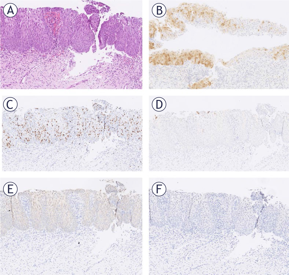
FIGURE 2.
Only case in the group with atypical squamous epithelium with patchy p16 expression and positive reaction for E4 human papillomavirus (HPV). The tissue was positive for HPV16 and 31 and the follow-up was negative. The same section at 100x magnification: (A) HE staining; (B) p16 staining, (C) Ki67 staining, (D) E4 HPV staining, (E) Nanog staining, F. SOX11 staining.
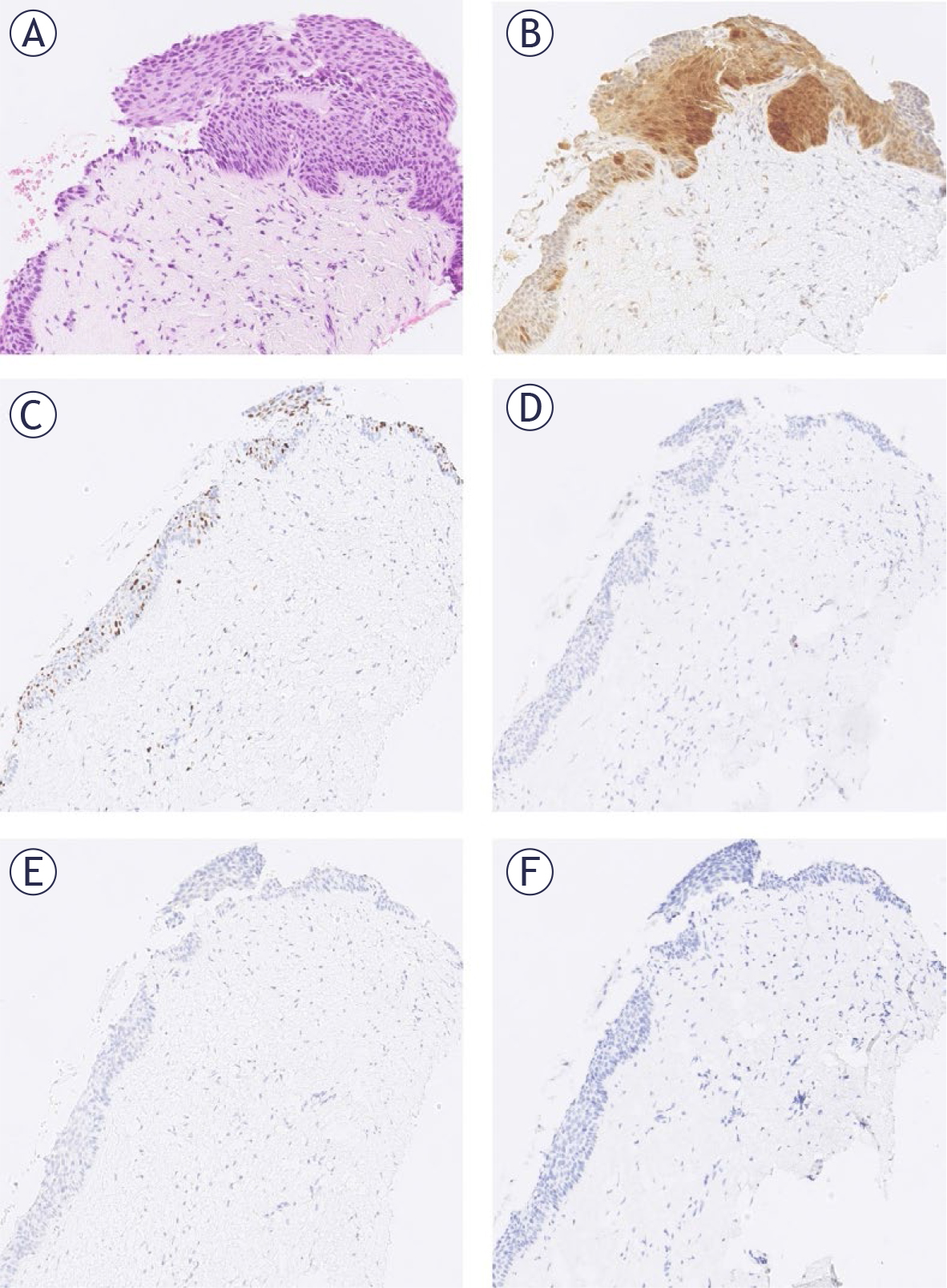
FIGURE 3.
One of the three cases in the group with atypical squamous epithelium with patchy p16 with CIN3 on follow-up. The tissue was positive for human papillomavirus (HPV) 52, 53,56, 66, 73. The same section at 100x magnification: (A) HE. (B) p16 staining. (C) Ki67 staining. (D) E4 HPV staining. (E) Nanog staining. (F) SOX11 staining.
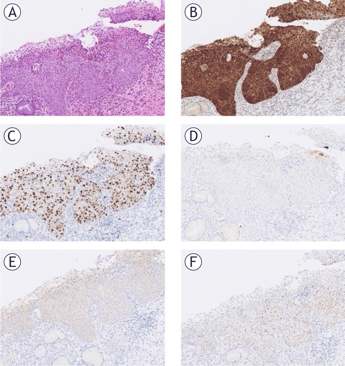
FIGURE 4.
Immunohistochemical reactions in the only cervical intraepithelial neoplasia 3 (CIN3) case with a positive SOX11 reaction in study. The same section at 100x magnification: (A) HE. (B) p16 staining. (C) Ki67 staining. (D) E4 HPV staining. (E) Nanog staining. (F) SOX11 staining.