![[18F]fluorocholine PET vs. [99mTc]sestamibi scintigraphy for detection and localization of hyperfunctioning parathyroid glands in patients with primary hyperparathyroidism: outcomes and resource efficiency Cover](https://paradigm-files.s3.eu-central-1.amazonaws.com/RAON/cover-image.jpg)
TABLE 1.
Workload of the profiles involved in the imaging process
| No. workers | SS + SPECT (hours) | No. patients/day | FCh PET (hours) | No. patients/day | |
|---|---|---|---|---|---|
| Pharmacist | 1 | 3 (12)* | 12 | ||
| RP preparation | 0.7 (2.8)* | 0,25 | |||
| Nurse | 1 | ||||
| Cannula placement | 0.75 (3)* | 3 | |||
| Cannula removal | 0.3 (1.2)* | 1,2 | |||
| Technologist | 2 | ||||
| RP application | 0.75 (3)* | 1,2 | |||
| Imaging time | 6 (24)* | 4 | |||
| QC dally test | 0.5 (2)* | 0,5 | |||
| Physician | 1 | ||||
| PH&CE and writing a report | 3 (12)* | 12 |

FIGURE 1.
Time workflow/scheme of radiopharmaceutical (RP) application and imaging of subtraction scintigraphy (SS) and single-photon emission tomography/computed tomography (SPECT/CT) in a working day. Approximate times are taken into account for all procedures.
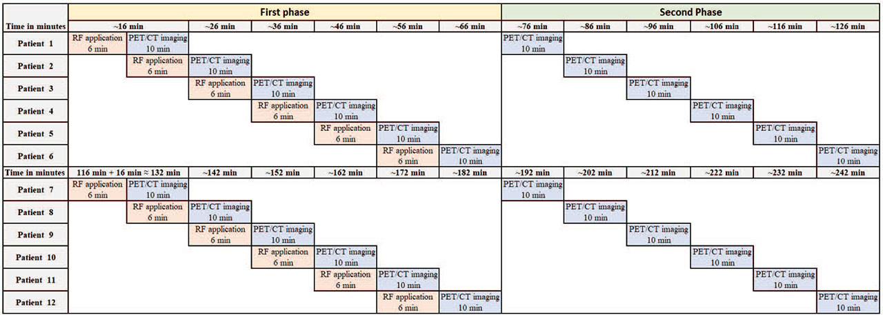
FIGURE 2.
Time workflow/scheme of radiopharmaceutical (RP) application and imaging of [18F]fluorocholine positron emission tomography (FCh-PET) in a working day. Approximate times are taken into account for all procedures.
TABLE 2.
The most common causes (percentages) of postoperative complications
| SS and SPECT/CT | FCh PET | |
|---|---|---|
| Tingling | 4/234 (1.7%) | 3/163 (1.8%) |
| Chvostek sign | 1/234 (0.4%) | 1/163 (0.6%) |
| Hungry bone syndrome | 1/234 (0.4%) | 2/163 (1.2%) |
| Malaise | 1/234 (0.4%) | N/C |
| Hoarseness | 1/234 (0.4%) | N/C |
| Postoperative Hypocalcemia | 1/234 (0.4%) | N/C |
| Hematoma | 1/234 (0.4%) | N/C |
| Deterioration of renal function | N/C | 1/163 (0.6%) |
| Reoperation | 29/234 (12.3%)* | 3/163 (1.8%)* |
| All | 39/234 (16.6%)** | 10/163 (6.1%)** |
TABLE 3.
The patient number and the required time of surgery expressed in minutes after subtraction scintigraphy (SS) and single-photon emission computed tomography/computed tomography (SPECT/CT) vs. [18F]fluorocholine positron emission tomography (FCh-PET)
| All pts | Mean | Median | SD | Min | Max | |
|---|---|---|---|---|---|---|
| MIBI SS and SPECT/CT | 234 | 67.37 | 60.00 | 36.88 | 20.00 | 280.00 |
| FCh-PET | 163 | 70.79 | 55.00 | 38.21 | 25.00 | 195.00 |
| p | 0.66 | |||||
| Solitary HPG | Mean | Median | SD | Min | Max | |
| MIBI SS and SPECT/CT | 195 | 63.56 | 50.00 | 33.04 | 20.00 | 235.00 |
| FCh-PET | 138 | 64.42 | 50.00 | 33.04 | 25.00 | 180.00 |
| p | 0.93 | |||||
| Multiple HPG | Mean | Median | SD | Min | Max | |
| MIBI SS and SPECT/CT | 39 | 94.26 | 100.00 | 43.65 | 25.00 | 235.00 |
| FCh-PET | 25 | 104.79 | 107.00 | 40.79 | 30.00 | 180.00 |
| p | 0.23 |
TABLE 4.
Influence of probable prognostic factors on overall survival (OS) and deasise-free survival (DFS)
| All pts | Mean | Median | SD | Min | Max | |
|---|---|---|---|---|---|---|
| No ioPTH | 123 | 60.00 | 50.00 | 33.91 | 25.00 | 195.00 |
| ioPTH | 39 | 103.00 | 100.00 | 31.27 | 40.00 | 195.00 |
| p | < 0.001 | |||||
| Solitary HPG | Mean | Median | SD | Min | Max | |
| No ioPTH | 113 | 57.30 | 50.00 | 31.05 | 25.00 | 180.00 |
| ioPTH | 25 | 96.60 | 90.00 | 29.71 | 40.00 | 155.00 |
| p | < 0.001 | |||||
| Multiple HPG | Mean | Median | SD | Min | Max | |
| No ioPTH | 10 | 85.55 | 70.00 | 49.90 | 30.00 | 195.00 |
| ioPTH | 15 | 116.33 | 110.00 | 30.49 | 80.00 | 195.00 |
| p | 0.03 |