
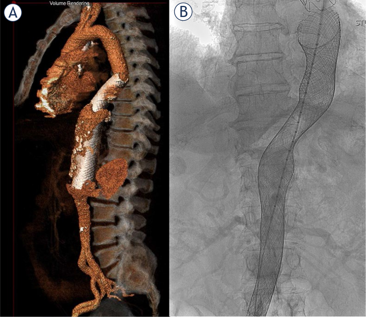
FIGURE 1.
Volume rendering of implanted multilayer flow modulators (MFMs) (A) and fluoroscopic image of implanted MFM (B) in thoracoabdominal aorta.
TABLE 1.
Age of patients at procedure, year of procedure, and location of multilayer flow modulator (MFM)
| Patient | Age | Procedure year | Location of MFM |
|---|---|---|---|
| 1. | 48 | 2012 | Thoracic aorta |
| 2. | 79 | 2012 | Thoracic aorta |
| 3. | 80 | 2012 | Thoracic aorta |
| 4. | 73 | 2013 | Aortic arch |
| 5. | 65 | 2013 | Thoracic aorta |
| 6. | 67 | 2016 | Aortic arch |
| 7. | 68 | 2016 | Thoracic aorta |
| 8. | 63 | 2016 | Abdominal aorta |
| 9. | 71 | 2016 | Thoracic aorta |
| 10. | 80 | 2017 | Abdominal aorta |
| 11. | 67 | 2017 | Aortic arch |
| 12. | 65 | 2017 | Aortic arch |
| 13. | 76 | 2018 | Thoracoabdominal aorta |
| 14. | 75 | 2018 | Thoracoabdominal aorta |
| 15. | 81 | 2018 | Abdominal aorta |
| 16. | 66 | 2018 | Abdominal aorta |
| 17. | 66 | 2019 | Thoracoabdominal aorta |
TABLE 2.
Time and cause of reinterventions after multilayer flow modulator (MFM) implantation
| Patient number | Time after MFM implantation (months) | Cause of reintervention; reintervention undertaken |
|---|---|---|
| 1. | 25 | Endoleak type I and enlargement of the aneurysm; implantation of another MFM |
| 2. | 3 | Insufficient proximal seal and collateral flow, resulting in enlargement of the aneurysm; implantation of another MFM |
| 3. | 1 | Insufficient proximal seal; implantation of another MFM |
| 4. | 8 | Enlargement of aneurysm and partial stenosis of subclavian artery; implantation of another MFM into previous MFM |
| 6. | 13 | Stenosis of brachiocephalic truncus; implantation of a stent, which resulted in a stroke |
| 9. | 9 | Migration of the MFM; implantation of another MFM |
| 10. | 74 | Displacement of MFM and stent graft; implantation of another stent graft |
TABLE 3.
Aneurysm size before and after multilayer flow modulator (MFM) implantation
| Patient number | Aneurysm volume (ml) | Aneurysm diameter (mm) | Difference between the last follow-up and before the MFM implantation | Duration of follow-up (months) | |||
|---|---|---|---|---|---|---|---|
| Before MFM implantation | At last follow-up | Before MFM implantation | At last follow-up | Volume (ml [%]) | Diameter (mm [%]) | ||
| 1. | 345 | 854 | 65 | 107 | 509 [147] | 42 [64] | 76 |
| 2. | 888 | 920 | 122 | 130 | 32 [4] | 8 [7] | 9 |
| 3. | 309 | 943 | 70 | 110 | 634 [205] | 40 [57] | 50 |
| 4. | 255 | 355 | 57 | 93 | 100 [39] | 36 [63] | 69 |
| 5. | 75 | 62 | 26 | 27 | −13 [−17] | 1 [4] | 14 |
| 6. | 197 | 330 | 44 | 76 | 133 [68] | 32 [73] | 49 |
| 7. | 343 | 549 | 65 | 85 | 206 [60] | 20 [31] | 28 |
| 8. | 311 | 266 | 48 | 68 | −45 [−14] | 20 [42] | 3 |
| 9. | 575 | 667 | 76 | 81 | 92 [16] | 5 [7] | 14 |
| 10. | 539 | 530 | 97 | 90 | −9 [−2] | −7 [−7] | 40 |
| 11. | 32 | 62 | 34 | 50 | 30 [94] | 16 [47] | 48 |
| 12. | 411 | 411 | 54 | 55 | 0 [0] | 1 [2] | 33 |
| 13. | 493 | 409 | 53 | 57 | −84 [−17] | 4 [8] | 24 |
| 14. | 262 | 332 | 60 | 75 | 70 [27] | 15 [25] | 23 |
| 15. | 307 | 223 | 59 | 81 | −84 [−27] | 22 [37] | 18 |
| 16. | 130 | 113 | 58 | 58 | −17 [−13] | 0 [0] | 19 |
| 17. | 250 | 250 | 54 | 60 | 0 [0] | 6 [11] | 7 |